Showing posts with label physiology. Show all posts

Friday, 19 September 2014

Facts about mitochondrial myopathies


Dear Friends:

if you are reading this booklet, it’s probably because you’ve just received a very bewildering diagnosis: mitochondrial myopathy. What is a mitochondrial myopathy, and what does the term mean? These are questions my wife, Jennifer, and I struggled with when our son, Michael, got his diagnosis in 1993.Mitochondrial myopathies have many different faces. As you will read in this booklet, dozens of varieties of mitochondrial diseases have been identified, with a complex array of symptoms. Some symptoms can be so mild that they’re hardly noticeable, while others are life-threatening.

Michael’s disease causes muscle weakness, muscle cramping, fatigue, lack of endurance and poor balance. You or your family member may have similar symptoms, yet each case is unique.

Continue reading on Muscular Dystrophy Association website.

Monday, 1 September 2014

Hormone Blues


A quick reference for recalling the hormones that should be memorized for the MCAT.. But in song form!

If we get views, we may make songs for a few more MCAT topics.

Thanks to Nathan Domek for both helping to record the song and filming/editing the actual video, and Morgan Komure for performing the song with me. 

Special thanks to The Snake, who portrayed a very convincing snake.

Sunday, 31 August 2014

The molecular bases of training adaptation


The Molecular Bases of Training Adaptation

Vernon G. Coffey and John A. Hawley
School of Medical Sciences, Exercise Metabolism Group, RMIT University, Melbourne, Victoria, Australia

Sports Med 2007; 37 (9): 737-763 REVIEW ARTICLE 0112-1642/07/0009-0737/$44.95/0 / 2007 Adis Data Information BV. All rights reserved.

Skeletal muscle is a malleable tissue capable of altering the type and amount of protein in response to disruptions to cellular homeostasis. The process of exercise induced adaptation in skeletal muscle involves a multitude of signalling mechanisms initiating replication of specific DNA genetic sequences, enabling subsequent translation of the genetic message and ultimately generating a series of amino acids that form new proteins. The functional consequences of these adaptations are determined by training volume, intensity and frequency, and the half-life of the protein. Moreover, many features of the training adaptation are specific to the type of stimulus, such as the mode of exercise. Prolonged endurance training elicits a variety of metabolic and morphological changes, including mitochondrial biogenesis, fast-to-slow fibre-type transformation and substrate metabolism. In contrast, heavy resistance exercise stimulates synthesis of contractile proteins responsible for muscle hypertrophy and increases in maximal contractile force output. Concomitant with the vastly different functional outcomes induced by these diverse exercise modes, the genetic and molecular mechanisms of adaptation are distinct. With recent advances in technology, it is now possible to study the effects of various training interventions on a variety of signalling proteins and early-response genes in skeletal muscle. Although it cannot presently be claimed that such scientific endeavours have influenced the training practices of elite athletes, these new and exciting technologies have provided insight into how current training techniques result in specific muscular adaptations, and may ultimately provide clues for future and novel training methodologies. Greater knowledge of the mechanisms and interaction of exercise-induced adaptive pathways in skeletal muscle is important for our understanding of the aetiology of disease, maintenance of metabolic and functional capacity with aging, and training for athletic performance. This article highlights the effects of exercise on molecular and genetic mechanisms of training adaptation in skeletal muscle.

Monday, 7 April 2014

Neuroscience Online, the Open-Access Neuroscience Electronic Textbook


Welcome to Neuroscience Online, the Open-Access Neuroscience Electronic Textbook.

This online, interactive courseware for the study of neuroscience is provided by the Department of Neurobiology and Anatomy at The University of Texas Medical School at Houston. The project is being developed under the direction of the Department Chair and Editor, John H. Byrne.

This content is intended to function as an online multimedia resource, and therefore is not supported as a downloadable or printable text. After you have taken the opportunity to explore this electronic textbook, please take a moment to participate in our brief user survey.

You have reached this version of Neuroscience Online by using an iOS or mobile device. Links with this HTML5 badge HTML5 have content optimized for iOS devices, however, not all animations are in HTML5. For best experience with NSO, we suggest use of a desktop computer with Flash capabilities.

Sunday, 9 March 2014

Exercise, how it can increase or decrease specific hormones and growth factors


A beatiful article on ScientificPhysicalTherapy.

Summary: The growth factor IGF-1 and the anabolic hormones growth hormone and testosterone stimulates protein synthesis and tissue regeneration. IGF-1 can be decreased by mild aerobic exercise at the lactate threshold which is approximately 50% VO2 max. This would be beneficial in cases where the patient has increased risk of cancer.

Exercising at levels higher than 60% VO2 max will increase IGF-1 and high resistance exercise will also increase IGF-1. Growth hormones are stimulated by high intensity exercise both short sprint exercise and resistance exercise if it provides high tissue stress.

Resistance exercises with high workloads are the most effective way to stimulate the release of testosterone. Clinically it may look like IGF-1, growth hormones and testosterone can only be stimulated later in the treatment program when the patient has progressed into higher resistance exercise protocols. It may however be possible for many patients even early on to stimulate the production of IGF-1 and the anabolic hormones by incorporating short interval maximal effort sprints on a cycle ergometer. This may be a way to increase the anabolic response and improve protein synthesis and tissue regeneration early on.

Tuesday, 18 February 2014

Nogo antibody study for spinal cord injury recovery


Project 

Nogo is a protein that only occurs in the central nervous system and prevents nerve regeneration. However, anti-Nogo antibodies make damaged nerve fibres grow.

As part of a worldwide study with pharmaceutical company Novartis, we are currently running tests involving paralysed patients.

Researchers 
N.N. 

FAQ on the Nogo antibody study

What is the Nogo antibody study actually about?

The study aims to prove that Nogo antibodies can make damaged nerve fibres regrow in humans (not just animals) and that this growth can lead to partial improvement of the body's functions and sensitivity. At present, this medication is in an early clinical stage and is undergoing pharmacokinetic tests and tests for tolerance (to determine possible side-effects). Treatment involves injecting Nogo antibodies into the spinal fluid and must be administered within 14 days of the accident. The Nogo antibody study is being conducted in Switzerland in close cooperation between the Paraplegic Centre at Balgrist University Hospital, various trauma centres and Novartis. We are the testing centre for Switzerland - our clinic is the only one in Switzerland offering this treatment at present.

The Nogo antibody study is giving people hope. Can people in the acute stage of spinal paralysis be helped?

Unfortunately, we will currently have to disappoint anyone who is hoping that this may be a cure. At best, we can expect improvements to movement and sensitivity, which will be useful to paralysed people in their day-to-day lives. For example, a tetraplegic may require less assistance or a paraplegic may regain some sensitivity in paralysed body parts. For the time being, the aim of the Nogo antibody study is to test tolerance in humans and check for any side-effects.

Read the rest here.

Saturday, 15 February 2014

Pathophysiology of the endocrine system


A wonderful link on Pathophysiology of the endocrine system by Colorado State University.

The pages in this book are initially categorized based on scientific discipline (e.g. Endocrine Pathophysiology, Medical Virology, etc.), but there are many links between disciplines (e.g. links between pages describing the physiology of the stomach in the Digestive Pathophysiology section and pages in the Endocrine Pathophysiology section describing gastric hormones.

How Do You Want to Grow?


Reviewed for medical accuracy by: Kestrson S., M.D.
Added to knowledge base: 10/16/08

Growth hormone supplementation has been widely publicized lately due to recent allegations against Jason Grimsley and David Segui of Major League Baseball fame.  People choose to take the supplement for a promise of augmented athletic performance or to turn back the clock on life, without chance of detection by governing agencies.  Before jumping on the bandwagon, you should know what growth hormone is and what its effects are.

What is Growth Hormone?

The polypeptide human growth hormone, hGH, is one of the most important naturally occurring hormones for normal growth to adult size.  It is normally secreted in a pulsatile fashion 6-12 times per day, meaning concentrations fluctuate throughout the day.  Growth hormone secretion peaks during puberty and decreases with age.  Secretion is increased by sleep, stress, starvation, puberty, exercise, hypoglycemia (low blood sugar), and other hormones (ie growth hormone releasing hormone).   

hGH secretion is inhibited by somatostatin (from the hypothalamus), somatomedins (hormones released by the hGH target organs in response to a rise in hGH), obesity, hyperglycemia (high blood sugar) and pregnancy.  It is important to understand that like many other hormone systems in the body, hGH and GHRH inhibit their own secretion as part of a negative feedback system.

What is the effect of Growth Hormone?

hGH is taken up by skeletal muscle and has many effects.  It acts directly to decrease glucose uptake into cells, increase hepatic gluconeogenisis, increase lipolysis (fat release from adipose cells), increase protein synthesis in muscle cells, increase lean body mass and increase release of insulin like growth factor (IGF-1) from the liver and locally in skeletal muscle.  IGF-1 increases bone length in children, increases protein synthesis in muscle and increases lean body mass.  Both growth hormone and IGF-1 are anabolic, non-androgenic compounds.  This means that it promotes protein synthesis without stimulating male secondary sex characteristics.

Are there diseases of Growth Hormone secretion?
Yes.  A deficiency in hGH can result in failure to grow and mild obesity.  Currently the FDA has approved growth hormone therapy in the treatment of childhood growth deficiencies and wasting diseases such as AIDS. 

There are also diseases of excess growth hormone secretion.  Excess hGH before puberty can cause gigantism and after puberty causes an increase in bone growth, organ size and glucose intolerance.

How can I increase my levels of hGH?
As already mentioned, exercise can naturally increase growth hormone secretion.  Research has shown that exercise counteracts the negative feedback mechanisms of GHRH and hGH allowing levels to rise unchecked by this feedback system.  Research has also shown that repeated exercise sessions led to an increase in hGH after each session and over a 24 hour period.  Results suggest that load and frequency of the resistance exercise are important determinants of hGH concentration while length of the workout is not. 

Over the short term, endurance exercise also increases growth hormone release.  Significant increases in plasma growth hormone was noted after 10 minutes of exercise above the lactic acid threshold (ie high intensity).  Multiple exercise sessions further increased the amount of growth hormone released per pulse rather than leading to feedback inhibition.  However, in response to chronic endurance exercise (greater than 3 weeks), some studies found a decreased growth hormone response to additional exercise bouts.  This suggests that the hGH response to endurance exercise is adaptive and may plateau with time.

Read the rest. It has nice information on GH supplements.

Friday, 17 January 2014

Anatomy drill and practice


Wiley

Wiley's Global Research business is a provider of content-enabled solutions to improve outcomes in research, education and professional practice with online tools, journals, books, databases, reference works and laboratory protocols.

This website offers amazing quizzes and sources to practise your knowledge on basic anatomy and physiology.

Tuesday, 6 August 2013

Antioxidants - too much of a good thing?


There is recently a lot of discussion done on the controversial effect of antioxidants on health. What is causing this discussion is the fact that the free radicals in our body (that were thought to be a danger for our health) are now found to play an important role in the defence mechanisms of it.

This article on medicalxpress.com discusses a new element on the effect of free radicals in our health. What is emerging is a new view that antioxidants are not a fix for everything, and that some degree of oxidant stress may be necessary for the body to work correctly.

Read it all - it is simple and very interesting.

Saturday, 3 August 2013

Mechanotransduction - converting mechanical stimulus to chemical activity


File:Cartilage forces.jpg

Why mobilization does good to our musculoskeletal system?

I wanted to understand the physiological mechanism behind this simple rule and I learned about mechanotransduction.

This is a biological phenomenon that we should be aware; the phenomenon of mechanotransduction - the mechanisms by which cells convert mechanical stimulus into chemical activity. This law can be and is applied in many physiological processes as well in many therapeutic techniques in Physical Therapy.

Below you can find an overview of the mechanism of mechanotransduction:

"The conversion of physical force into biochemical information is fundamental to development and physiology. It provides a simple means by which cells and organisms can ensure structural stability, as well as a way to regulate morphogenetic movements to generate precise three-dimensional structures. In the vascular system, pressure and shear stress from pumping blood influence the morphology and pathology of the heart and vasculature. Bone is shaped by forces from gravity and muscle contraction. Hearing and touch are based on neural responses to pressure. Inflation and deflation of the lungs regulate their physiology. Coordinated growth of tissues is guided by mechanical forces, and failure of these mechanisms contributes to cancer. Mechanosensitivity in one form or another appears to be a property shared by all cells of the body and by all phyla from mammals to bacteria".

Friday, 5 July 2013

Pelvic floor muscles physiology


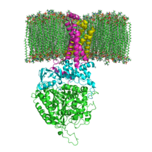
Succinate Dehydrogenase 1YQ3 and Membrane.png

I recently read an research article on pelvic floor muscle assessment and I found interesting the following parts:
  1. The pelvic floor muscles (PFM) consist of approximately 70% slow-twitch (type 1) and 30% fast-twitch (type 2) muscle fibres (Gilpin et al, 1989).
  2. During a vaginal examination using the distal pad of the index finger to palpate the perivaginal muscles, a definite bulging and lifting of the muscles are felt during a contraction, and in both the contracted and relaxed state, areas of atrophy can be detected.
  3. A moderate to strong contraction of the levator ani muscles has both a squeeze and a lift component.
  4. Regarding different muscle components (that is, fast- and slow-twitch fibres), it has been shown that fast fibres are recruited only during activities involving speed and/or power (Edwards, 1978) and if the response to a command to contract the PFM maximally is sluggish, then it is probable that only slow-twitch muscle fibres are being used. Jones and Rutherford (1987) reported that in some untrained muscles, the fast-firing muscle fibres are never recruited, and so these may need targeting in some other way during PFM rehabilitation. Gosling et al (1981) described the PFM slow-twitch fibres as being responsible for maintaining continuous muscle activity over prolonged periods, with the fast-twitch fibres recruited reflexly during sudden increases in intra-abdominal pressure, for example when coughing. A reduction in slow-twitch activity would therefore manifest itself in a short duration contraction and few repetitions, and PFM in this category may thus be responsible for decreased support of the proximal urethra and reduced urethral occlusion. A reduction in fast-twitch fibre activity may result in a slower, weaker reflex response to increases in intra-abdominal pressure. Logically, one should assess both the fast- and slow-twitch muscle fibre activity before planning an exercise regimen.
  5. Exercise programme planning: the plethora of PFM exercise regimens in the literature indicates a lack of standardisation and, when compared with general rehabilitation methodology, the reports show lack of scientific application. There is a wide range of muscular strength and endurance across any female population, and so it is postulated that a uniform, standard regimen is not appropriate. Instead, assessment of the fast- and slow-twitch components, leading to an individual exercise programme, has been proposed, to target specifically the weakness of each individual patient.
  6. Muscle weakness leads to reduction in mitochondrial oxidative capacity as indicated by a decrease in the succinate dehydrogenase (SDH) activity (Eriksson and Haggmark, 1979). Consequently, these same authors maintain that it is necessary to gradually increase the SDH reserves by repeated contractions which will deplete levels and stimulate greater production. This reinforces the theory of overload and supports the need for a programme of regular daily contractions advanced in this study.

Interesting also to know that SDH or succinate-coenzyme Q reductase (SQR) or respiratory Complex II is an enzyme complex, bound to the inner mitochondrial membrane of mammalian mitochondria and many bacterial cells. It is the only enzyme that participates in both the citric acid cycle and the electron transport chain.

The fundamental role of SDH in the electron transfer chain of mitochondria makes it vital in most multicellular organisms, removal of this enzyme from the genome has also been shown to be lethal at the embryonic stage in mice. Mutations of different genes used in its construction can lead to:
  • Leigh syndrome, mitochondrial encephalopathy, and optic atrophy
  • Tumorogenesis in chromaffin cells, causing hereditary paraganglioma and hereditary pheochromocytoma. Tumors tend to be malignant. It can also lead to decreased life-span and increased production of superoxide ions.
  • Decreased life-span, increased production of superoxide ions, hereditary paraganglioma and hereditary pheochromocytoma. Tumors tend to be benign. These mutations are uncommon.
  • Hereditary paraganglioma and hereditary pheochromocytoma. Tumors tend to be benign, and occur often in the head and neck regions. These mutations can also decrease life-span and increase production of superoxide ions.

Mammilian succinate dehydrogenase functions not only in mitochondrial energy generation, but also has a role in oxygen sensing and tumor suppression; and, therefore, is the object of ongoing research.

Wednesday, 3 July 2013

Golgi tendon reflex vs. stretch reflex


Two different reflexes exist in skeletal muscles: the Golgi tendon reflex and the stretch reflex. It is very important for a Physical Therapist to understand the structure and function of these two reflexes, because he will use their effect on muscles in many different cases.

In the conceptual model above, I have drawn an agonist muscle (the big orange cylinder), an antagonist muscle (the smaller orange cylinder) and a spinal segment (the grey region). In the agonist muscle I have also drawn two sensory organs: the Golgi tendon organ and the muscle spindle.

The Golgi tendon organ senses changes in muscle tension. It is a proprioceptive sensory receptor organ that is located at the origins and insertion of skeletal muscle fibers into the tendons of skeletal muscle. It provides the sensory component of the Golgi tendon reflex. In a Golgi tendon reflex, skeletal muscle contraction causes the agonist muscle to simultaneously lengthen and relax. Though muscle tension is increasing during the contraction, alpha motor neurons in the spinal cord supplying the muscle are inhibited through the Inhibitory Postsynaptic Potential. However, antagonistic muscles are activated.

An inhibitory postsynaptic potential (IPSP) is a kind of synaptic potential that makes a postsynaptic neuron less likely to generate an action potential. The opposite of an inhibitory postsynaptic potential is an excitatory postsynaptic potential (EPSP), which is a synaptic potential that makes a postsynaptic neuron more likely to generate an action potential. They can take place at all chemical synapses which use the secretion of neurotransmitters to create cell to cell signalling. Inhibitory presynaptic neurons release neurotransmitters which then bind to the postsynaptic receptors; this induces a postsynaptic conductance change as ion channels open or close. An electrical current is generated which changes the postsynaptic membrane potential to create a more negative postsynaptic potential.

Muscle spindles are sensory receptors within the belly of a muscle, which primarily detect changes in the length of this muscle. They convey length information to the central nervous system via sensory neurons. This information can be processed by the brain to determine the position of body parts. The responses of muscle spindles to changes in length also play an important role in regulating the contraction of muscles, by activating motoneurons via the stretch reflex to resist muscle stretch. The stretch reflex (myotatic reflex) is a muscle contraction in response to stretching within the muscle. It is a monosynaptic reflex which provides automatic regulation of skeletal muscle length.

When a muscle lengthens, the muscle spindle is stretched and its nerve activity increases. This increases alpha motor neuron activity, causing the muscle fibers to contract and thus resist the stretching. A secondary set of neurons also causes the opposing muscle to relax. The reflex functions to maintain the muscle at a constant length.

Wednesday, 26 June 2013

Mechanisms of inflammatory pain


By B. L. Kidd1 and L. A. Urban

One of the cardinal features of inflammatory states is that normally innocuous stimuli produce pain. Since the publication of the Melzack–Wall gate control theory in 1965,45 it has been widely appreciated that the nervous system exhibits a range of responses according to different conditions (‘neural plasticity’). Subsequent research has characterized the mechanisms by which these changes occur and highlighted the importance of environmental factors on perception of pain.

This review focuses on key peripheral mechanisms that result in the hypersensitivity state that accompanies inflammation. Recent studies are described which characterize a series of receptors, ion channels and transmitters involved in inflammatory pain. The mechanisms by which inflammatory mediators interact with neurones to produce hypersensitivity are also explored.

Read more.

Temperature regulation during exercise - Bikram Yoga or not?


Somebody told me yesterday about the concept of Bikram Yoga. I am not an expert and I do not want to discourage people that support it, but, based on my knowledge, I believe that the side effects and the risks of it outweigh the possible benefits. The main reasons I believe that are the following:

  1. Exhaustion of body temperature control mechanisms
  2. Unnecessary heart overload
  3. Unhealthy temperature for the skin and hyperthermia
  4. Oversweating and dehydration
  5. Possible instability of joints due to excess mobility under high temperature
So, I thought it would be nice to post an article on some basic temperature regulation principles. A collection of sources on how temperature is regulated during exercise and the effects of it on the body mechanisms follows.

As in other mammals, thermoregulation is an important aspect of human homeostasis. Most body heat is generated in the deep organs, especially the liver, brain, and heart, and in contraction of skeletal muscles. Humans have been able to adapt to a great diversity of climates, including hot humid and hot arid. High temperatures pose serious stresses for the human body, placing it in great danger of injury or even death. For humans, adaptation to varying climatic conditions includes both physiological mechanisms resulting from evolution and behavioural mechanisms resulting from conscious cultural adaptations.


In hot conditions

  • Eccrine sweat glands under the skin secrete sweat (a fluid containing mostly water with some dissolved ions) which travels up the sweat duct, through the sweat pore and onto the surface of the skin. This causes heat loss via evaporative cooling; however, a lot of essential water is lost.
  • The hairs on the skin lie flat, preventing heat from being trapped by the layer of still air between the hairs. This is caused by tiny muscles under the surface of the skin called arrector pili muscles relaxing so that their attached hair follicles are not erect. These flat hairs increase the flow of air next to the skin increasing heat loss by convection. When environmental temperature is above core body temperature, sweating is the only physiological way for humans to lose heat.
  • Arteriolar vasodilation occurs. The smooth muscle walls of the arterioles relax allowing increased blood flow through the artery. This redirects blood into the superficial capillaries in the skin increasing heat loss by convection and conduction.


In cold conditions

  • Sweat stops being produced.
  • The minute muscles under the surface of the skin called arrector pili muscles (attached to an individual hair follicle) contract (piloerection), lifting the hair follicle upright. This makes the hairs stand on end which acts as an insulating layer, trapping heat. This is what also causes goose bumps since humans don't have very much hair and the contracted muscles can easily be seen.
  • Arterioles carrying blood to superficial capillaries under the surface of the skin can shrink (constrict), thereby rerouting blood away from the skin and towards the warmer core of the body. This prevents blood from losing heat to the surroundings and also prevents the core temperature dropping further. This process is called vasoconstriction. It is impossible to prevent all heat loss from the blood, only to reduce it. In extremely cold conditions excessive vasoconstriction leads to numbness and pale skin. Frostbite only occurs when water within the cells begins to freeze, this destroys the cell causing damage.
  • Muscles can also receive messages from the thermo-regulatory center of the brain (the hypothalamus) to cause shivering. This increases heat production as respiration is an exothermic reaction in muscle cells. Shivering is more effective than exercise at producing heat because the animal remains still. This means that less heat is lost to the environment via convection. There are two types of shivering: low intensity and high intensity. During low intensity shivering animals shiver constantly at a low level for months during cold conditions. During high intensity shivering animals shiver violently for a relatively short time. Both processes consume energy although high intensity shivering uses glucose as a fuel source and low intensity tends to use fats. This is a primary reason why animals store up food in the winter.[citation needed]
  • Mitochondria can convert fat directly into heat energy, increasing the temperature of all cells in the body. Brown fat is specialized for this purpose, and is abundant in newborns and animals that hibernate.

What is happening during exercise?

During strenuous exercise the body's heat production may exceed 1000 W. Some of the heat produced is stored, raising body core temperature by a few degrees. Rises in body temperature are sensed by central and skin thermoreceptors and this sensory information is processed by the hypothalamus to trigger appropriate effector responses. Other sensory inputs from baroreceptors and osmoreceptors can modify these responses. Evaporation of sweat and increased skin blood flow are effective mechanisms for the dissipation of heat from the body but dehydration impairs the capacity to sweat and lose body heat. Hot, humid environments or inappropriate clothing may compromise the ability to lose heat from the body. Exercise training improves tolerance to exercise in the heat by increasing the sensitivity of the sweat rate/core temperature relationship, decreasing the core temperature threshold for sweating and increasing total blood volume.


How does cold temperature affect heart rate?

Unless you are starting to dye or get severe hypothermia your blood pressure increases. Because your muscles are tensing to confiscate for the cold weather. thats why you shiver. Since your body is doing that your organs demand for more blood and since your body works together the heart pumps out more blood to fulfill the need, therefore increasing your blood pressure.


How does hot temperature affect heart rate?

The blood vessels of the body, particularly the peripheral blood vessels, i.e those of the skin; can dilate or contract depending on body temperature. When the temperature of the body drops, the peripheral blood vessels contract to conserve heat. When the body's temperature increases above normal, the peripheral blood vessels dilate to allow more heat to escape. The blood vessels dilating lowers the blood pressure in the body, because the blood distribution has increased to areas which previously had less distribution. The body now has to compensate, because of the change in pressure less blood is being pumped to the vital organs of the body. It does this by increasing the cardiac output, the amount of blood pumped out of the heart per given time. Cardiac output can be increased by increasing the heart rate. Thus an increase in temperature, through the vasodilation of blood vessels causes a drop in blood pressure, which is compensated for by increasing the cardiac output, which can be achieved through increasing the heart rate. Therefore heart rate increases when temperature increases.


How does sweating help us to cool down?

When we sweat, our skin and clothing become covered with water. If the atmospheric humidity is low, this water evaporates easily. The heat energy needed to evaporate the water comes from our bodies. So this evaporation cools our bodies, which have too much heat. For the same reason splashing water on ourselves when it is hot feels good. Being wet during cold weather, however can excessively chill us because of this same evaporation effect.

Sweating is therefore the human body's primary cooling mechanism. Because this mechanism uses water, we need to replace lost fluids by drinking more fluids in hot weather. This is especially true after exercising or working in hot weather.


Does sweating help you lose weight?

Sweat happens. Throughout every day of your life, you exude a watery blend of urea, salts, sugars and ammonia from your pores. Most people presume that when you sweat, you lose weight. It's true that when you sweat, you shed some weight. However, most of the weight you lose is water weight, and you regain that as soon as you rehydrate. Any real weight loss that occurs when you sweat happens because of what you are doing that makes you sweat, and not because of the sweating.


Does sweat remove toxins from the body?

Toxins in the body include heavy metals and various chemicals such as pesticides, pollutants, and food additives. Drugs and alcohol have toxic effects in the body. Toxins are produced as normal by-products in the intestines by the bacteria that break down food. The digestion of protein also creates toxic by-products in the body.

The body has natural methods of detoxification. Individual cells get detoxified in the lymph and circulatory system. The liver is the principle organ of detoxification, assisted by the kidneys and intestines. Toxins can be excreted from the body by the kidneys, bowels, skin, and lungs. Detoxification treatments become necessary when the body's natural detoxification systems become overwhelmed. This can be caused by long-term effects of improper diet, stress, overeating, sedentary lifestyles, illness, and poor health habits in general.

Sweat does contain trace amounts of toxins but definitely won't help clear the body of mercury or other metals. Almost all toxic metals in the body are excreted through urine or feces. Less than 1% are lost through sweat. Sweating for the sake of sweating (e.g. sauna "treatments") has no benefits. In fact, heavy sweating can impair your body's natural detoxification system due to dehydration.

Sources:

Sunday, 23 June 2013

The circadian rhythm


A circadian rhythm is any biological process that displays an endogenous, entrainable oscillation of about 24 hours. These rhythms are driven by a circadian clock, and rhythms have been widely observed in plants, animals, fungi and cyanobacteria. The term circadian comes from the Latin circa, meaning "around" (or "approximately"), and diem or dies, meaning "day". The formal study of biological temporal rhythms, such as daily, tidal, weekly, seasonal, and annual rhythms, is called chronobiology. Although circadian rhythms are endogenous ("built-in", self-sustained), they are adjusted (entrained) to the local environment by external cues called zeitgebers, commonly the most important of which is daylight.

Sunday, 16 June 2013

The effects of overtraining


I found myself extremely tired these days due to overtraining. So, I was looking ιν μυ βοοκσ ανδ online to find some information of the effects and the pathophysiology of overtraining. I am just pasting some things from Wikipedia that I find important and interesting for me as a summary of the things I need to always remember for myself and my future patients:

Overtraining is a physical, behavioral, and emotional condition that occurs when the volume and intensity of an individual's exercise exceeds their recovery capacity. They cease making progress, and can even begin to lose strength and fitness. Overtraining is a common problem in weight training, but it can also be experienced by runners and other athletes.

Like pharmacological drugs, physical exercise may be chemically addictive. One theory is that this addiction is due to natural endorphins and dopamine generated and regulated by the exercise. Whether strictly due to this chemical by-product or not, some people can be said to become addicted to or fixated on psychological/physical effects of physical exercise and fitness. This may lead to overexercise, resulting in the "overtraining" syndrome.

Physiology

Ιmprovements in strength and fitness occur only after the rest period following hard training (see supercompensation). This process can take days to complete, depending on the intensity and duration of exercise leading to the overtrained state. If sufficient rest is not available, then complete regeneration cannot occur. If this imbalance between excess training and inadequate rest persists, then the individual's performance will eventually plateau and decline. Mild over training may require several days of rest or reduced activity to fully restore an athlete's fitness. If prompt attention is not given to the developing state and an athlete continues to train and accumulate fatigue, the condition may come to persist for weeks.

Overtraining occurs more readily if the individual is simultaneously exposed to other physical and psychological stressors, such as jet lag, ongoing illness, overwork, menstruation, poor nutrition etc. It is a particular problem for bodybuilders and other dieters who engage in intense exercise while limiting their food intake.

A number of possible mechanisms for overtraining have been proposed:
  • Microtrauma to the muscles are created faster than the body can heal them.
  • Amino acids are used up faster than they are supplied in the diet. This is sometimes called "protein deficiency".
  • The body becomes calorie-deficient and the rate of break down of muscle tissue increases.
  • Levels of cortisol (the "stress" hormone) are elevated for long periods of time.
  • The body spends more time in a catabolic state than an anabolic state (perhaps as a result of elevated cortisol levels).
  • Excessive strain to the nervous system during training.
Overtraining may be accompanied by one or more concomitant symptoms:
  • Persistent muscle soreness
  • Persistent fatigue
  • Elevated resting heart rate
  • Reduced heart rate variability
  • Increased susceptibility to infections
  • Increased incidence of injuries
  • Irritability
  • Depression
  • Mental breakdown
Treatment

Allowing more time for the body to recover:
  • Taking a break from training to allow time for recovery.
  • Reducing the volume and/or the intensity of the training.
  • Suitable periodization of training.
  • Splitting the training program so that different sets of muscles are worked on different days.
  • Increase sleep time.
  • Deep-tissue or sports massage of the affected muscles.
  • Self-massage or rub down of the affected muscles.
  • Cryotherapy and thermotherapy.
  • Temperature contrast therapy (contrast showers etc.).
Changing diet:
  • Ensuring that calorie intake at least matches expenditure.
  • Ensuring total calories are from a suitable macronutrient ratio.
  • Addressing vitamin deficiencies with nutritional supplements.

Saturday, 15 June 2013

Muscular dystrophy


Muscular dystrophy is one of the most frequently encountered genetic diseases, affecting an estimated 1 in every 3500 males (but many fewer females). It is associated with the progressive degeneration of skeletal and cardiac muscle fibers, weakening the muscles and leading ultimately to death from respiratory or cardian failure.

Muscular dystrophies are caused are caused by the absence or defect of one or more proteins that make up the costameres in striated muscles. Costameres are clusteres of structural and regulatory proteins that link the Z disks of the outermost myofibrils to the sarcolemma and extracellular matrix. Proteins of the costameres serve multiple roles, including lateral transmission of force from the sarcomeres to the extracellular matrix and neighboring muscle fibers, stabilization of the sarcolemma against physical forces during muscle fiber contraction or stretch, and initiation of intracellular signals that link contractile activity with regulation of muscle cell remodeling. Defects in a number of specific costamere proteins have been demonstrated to cause various types of muscular dystrophy.

Duchenne muscular dystrophy is a sex-linked recessive disorder caused by a defect in a gene on the X chromosome that codes for the protein dystrophin. It is more common in males since they possess only 1 X chromosome, which increases the possibility of the expression of the defected gene. The defected gene can result in either a nonfunctional or missing protein leading to muscle fibers susceptibility to membrane rupture and death. The condition therefore progresses with muscle use and age. Symptoms of weakness in the muscles of the hip girdle and trunk become evident at about 2 to 6 years of age and most affected individuals do not survive far beyond the age of 20. Preliminary attempts are being made to treat the disease by inserting the normal gene into dystrophic muscle cells.

The other major forms of Muscular Dystrophy are Becker, limb-girdle, congenital, facioscapulohumeral, myotonic, oculopharyngeal, distal, and Emery-Dreifuss muscular dystrophy.

References:

  • National Institutes of Health: Department of  Health and Human Services. Report to Congress on Implementation of the Muscular Dystrophy Community Assistance. c2006 [cited 2006 May]. URL: http://www.ninds.nih.gov/find_people/groups/mdcc/md_care_implementation.pdf.
  • Widmaier EP, Raff H, Strang KT. Vander's Human Physiology: the mechanisms of body function. 12 ed. New York: McGraw-Hill International Edition; 2011.