Sunday 30 June 2013

Physical Therapy for Multiple Sclerosis


Multiple sclerosis (MS) is an inflammatory autoimmune disease in which myelin sheaths around nerve cells of the brain and spinal cord are damaged, leading to loss of myelin and scarring. When this nerve covering is damaged, nerve signals slow down or stop. The nerve damage is caused by inflammation. Inflammation occurs when the body's own immune cells attack the nervous system. This can occur along any area of the brain, optic nerve, and spinal cord.

MS affects women more than men. The disorder is most commonly diagnosed between ages 20 and 40, but can be seen at any age. It is unknown what exactly causes this to happen. The most common thought is that a virus or gene defect, or both, are to blame. Environmental factors may play a role. You are slightly more likely to get this condition if you have a family history of MS or live in an part of the world where MS is more common.

Symptoms

Symptoms vary, because the location and severity of each attack can be different. Episodes can last for days, weeks, or months. These episodes alternate with periods of reduced or no symptoms (remissions). Fever, hot baths, sun exposure, and stress can trigger or worsen attacks. Heat intolerance in MS shows up as a "pseudoexacerbation" - the experience of having symptoms appear or worsen due to heat exposure. This is different than a true relapse. In the case of a pseudoexacerbation, when the body’s temperature returns to normal, these symptoms disappear. No damage, such as inflammation, demyelination or new lesions, has been done during these pseudoexacerbations.

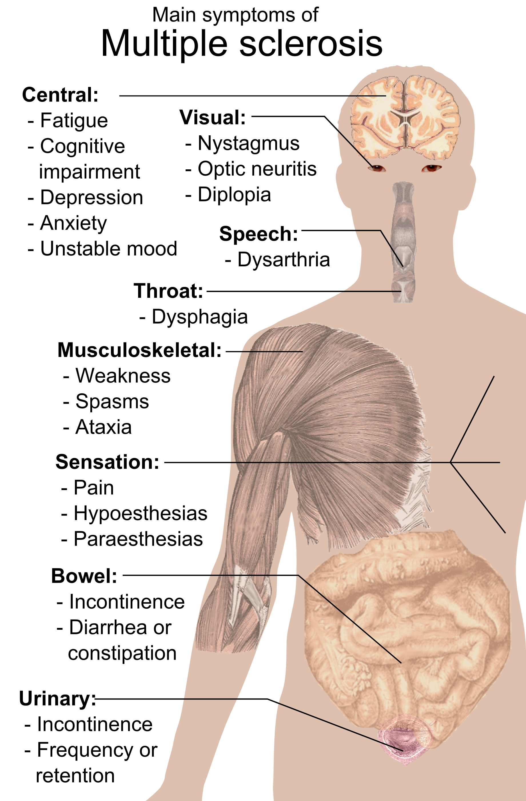
It is common for the disease to return (relapse). However, the disease may continue to get worse without periods of remission. Because nerves in any part of the brain or spinal cord may be damaged, patients with multiple sclerosis can have symptoms in many parts of the body - see the picture above for the main symptoms.

How does heat affect MS?

Demyelination slows the ability of the nerves to function, and heat further slows down nerve impulse transmission in demylinated regions. Even a very slight increase of as little as one-quarter to one-half a degree in the body’s core temperature is enough to cause symptoms of heat intolerance.

Multiple sclerosis is a demyelinating disease of the central nervous system, characterized by a relapsing and remitting or, alternatively, by a steady and continuous course of dysfunction. The hallmark of the disease is heterogeneity. How the disease presents itself depends on the exact location and total accumulation of lesions and on the impact of demyelination on nerve conduction. Conduction maybe be enhanced, producing positive symptoms like tic douloureux, paresthesia, or tingling, or conduction may be diminished, producing negative symptoms like blindness and paralysis. Diminished conduction may be total or partial, with decreases in the maximum of frequency or velocity of conduction. During remission, which may last months or years, symptoms improve as inflammation subsides, sodium channels migrate into the bare axon, remyelination of the denuded site occurs, and, ultimately, conduction resumes through the lesion sites. The deficit may also become permanent as axons degenerate. During shorter periods lasting hours or days, symptoms may also worsen, as conduction through old lesions fluctuates; conduction is “highly insecure” and subject to inflammatory factors like nitric oxide and temperature changes.

Heat worsens and cooling improves negative symptoms of multiple sclerosis, sometimes dramatically so. The underlying mechanism relates to the influence of temperature on sodium channels and on current necessary for depolarization of the axon. Increases in temperature diminish the depolarizing current, whereas decreases in temperature have the opposite effect. Sensitivity can be extreme, and very small changes can have profound effects. Heating, for example by radiation from the sun, can turn a limping gait into no gait at all. Alternatively, hot air from a hair dryer can turn a hopeful morning into an exhausting one. Cooling, on the other hand, by sitting in a cool bath or shower, can turn the “disease off” (at least for a little while) and give an individual back the freedom for exercise or work.

A University of Michigan researcher, Dr. Joseph Cannon, studied the effects of exercise on the immune system in rats and humans. He noted that the immune system may become more active in any situation when there’s a rise in body temperature (such as during strenuous activity); in other words, that exercise can have the same effect on the immune system as fever does. Since the immune system takes its cue from a rise in body temperature during illness, it evidently does the same when any rise in temperature occurs, such as during strenuous exercise, a hot day, a hot bath, or emotional stress.

Any increase in temperature, then, could result in a situation where white blood cells may be produced to fight an enemy—with no enemies around to fight! Imagine some of these white blood cells entering the CNS, gathering at various points along the nerves. These gatherings of white blood cells result in inflammation, or swelling, resulting in pressure, or a “squeeze” on nerve cells in the area. Depending on which nerve cells are being squeezed, new symptoms may appear in that part of the body associated with those nerve cells.

Diagnosis

Symptoms of MS may mimic those of many other nervous system disorders. The disease is diagnosed by ruling out other conditions. People who have a form of MS called relapsing-remitting may have a history of at least two attacks, separated by a period of reduced or no symptoms.

The health care provider may suspect MS if there are decreases in the function of two different parts of the central nervous system (such as abnormal reflexes) at two different times.

A neurological exam may show reduced nerve function in one area of the body, or spread over many parts of the body. This may include:
  • Abnormal nerve reflexes
  • Decreased ability to move a part of the body
  • Decreased or abnormal sensation
  • Other loss of nervous system functions
An eye examination may show:
  • Abnormal pupil responses
  • Changes in the visual fields or eye movements
  • Decreased visual acuity
  • Problems with the inside parts of the eye
  • Rapid eye movements triggered when the eye moves
Tests to diagnose multiple sclerosis include:
  • Lumbar puncture (spinal tap) for cerebrospinal fluid tests, including CSF oligoclonal banding
  • MRI scan of the brain and MRI scan of the spine are important to help diagnose and follow MS
  • Nerve function study (evoked potential test)

Treatment

There is no known cure for multiple sclerosis at this time. However, there are therapies that may:
  1. slow the disease through medication
  2. treat exacerbations (also called attacks, relapses, or flare-ups) by reducing inflammation through corticosteroids
  3. manage symptoms
  4. improve function and safety through physical therapy
  5. provide emotional support
In combination, these treatments enhance the quality of life for people living with MS.

Are there alternative therapies for MS?

Complementary and alternative medicine (CAM) includes a variety of interventions - from exercise and dietary supplements to stress management strategies, biofeedback, and acupuncture. These therapies - which come from many different disciplines and traditions - are generally considered to be outside the realm of conventional medicine. When used in combination with conventional medicine, they are referred to as “complementary;” when used instead of conventional medicine, they are referred to as “alternative.” In the United States today, approximately 75% of people with MS use one form or another of CAM, generally in combination with their prescribed MS treatments.

Physical Therapy and MS

The role of rahabilitation in managing MS:
  • Physical therapy, speech therapy, occupational therapy, and support groups
  • Assistive devices, such as wheelchairs, bed lifts, shower chairs, walkers, and wall bars
  • A planned exercise program early in the course of the disorder
  • A healthy lifestyle, with good nutrition and enough rest and relaxation
  • Avoiding fatigue, stress, temperature extremes, and illness
  • Changes in what you eat or drink if there are swallowing problems
  • Making changes around the home to prevent falls
  • Social workers or other counseling services to help you cope with the disorder and get assistance (such as Meals-on-Wheels)
Physical Therapy cannot treat the primary symptoms of MS, but can do a lot of things for the secondary effects that are the result of them. The Physical Therapist evaluates and addresses the body’s ability to move and function, with particular emphasis on walking, strength, balance, posture, fatigue, and pain. PT might include stretching, range-of-motion and strengthening exercises, gait training, and training in the use of mobility aids (canes, crutches, scooters and wheelchairs) and other assistive devices.

The ultimate goal is to achieve and maintain optimal functioning and prevent unnecessary complications such as de-conditioning, muscle weakness from lack of mobility, and muscle contractures related to spasticity. Building a treatment plan, a Physical Therapist should follow the WHO's ICF model and base his strategy based on that. I have already posted an article about a beautiful tool one can use in order to build his strategy - the getptsmart.com. Regarding body structures and functions, a Physical Therapist should evaluate and manage:
  1. Pain
  2. Fatigue and fitness level
  3. Balance problems and poor coordination
  4. Muscle strength
  5. Bladder and bowel control
Taking into consideration the personal and environmental factors of the specific patient together with the specific activities and participation limitations will help the Physical Therapist build a successful program tailored to the patient he is dealing with. Patient and family education always plays a major role in the rehabilitation process.

Some general techniques used in Physical Therapy are the following: 
Furthermore, taking advantage of the cooling properties of water may help attenuate the consequences of heat sensitivity. In a study done by White et al. (2000), exercise pre-cooling via lower body immersion in water of 16-17°C for 30 minutes allowed heat sensitive individuals with MS to exercise in greater comfort and with fewer side effects by minimizing body temperature increases during exercise. Hydrotherapy exercise in moderately cool water of 27-29°C water can also be advantageous to individuals with MS. Temperatures lower than 27°C are not recommended because of the increased risk of invoking spasticity.

Here is a a Physical Therapy case study that uses the getptsmart.com tool. I find it amazing.


Sources:

We Can! - ways to enhance children's activity & nutrition


Boy raising arms in the air

National Heart Lung and Blood Institute Logo

A great movement of the National Heart, Lung, and Blood Institute in the USA, We Can! (Ways to Enhance Children's Activity & Nutrition) is a national movement designed to give parents, caregivers, and entire communities a way to help children 8 to 13 years old stay at a healthy weight.

Research shows that parents and caregivers are the primary influence on this age group. The We Can! national education program provides parents and caregivers with tools, fun activities, and more to help them encourage healthy eating, increased physical activity, and reduced time sitting in front of the screen (TV or computer) in their entire family.

We Can! also offers organizations, community groups, and health professionals a centralized resource to promote a healthy weight in youth through community outreach, partnership development, and media activities that can be adapted to meet the needs of diverse populations. Science-based educational programs, support materials, training opportunities, and other resources are available to support programming for youth, parents, and families in the community.

Saturday 29 June 2013

Foot drop: L4/5 root or peroneal nerve?


Foot drop is a gait abnormality in which the dropping of the forefoot happens due to weakness, damage to the peroneal nerve or paralysis of the muscles in the anterior portion of the lower leg.

Possible lesion sites causing foot drop include (going from peripheral to central):
  • Peroneal nerve - common, deep or superficial;
  • Sciatic nerve;
  • Lumbosacral plexus;
  • L5 nerve root - herniated disc;
  • Spinal cord - poliomyelitis, tumor;
  • Brain (uncommon, but often overlooked) - stroke, tumor;

Other causes of foot drop are diabetes, trauma, motor neuron disease (MND), adverse reaction to a drug or alcohol, neuromuscular disease, and multiple sclerosis.

The challenge for a Physical Therapist is to differentiate between the orthopedic causes; in other words, to find the location of entrapment or trauma. Is it distally or proximally to the spinal cord? The diagnosis of diabetes, MND, and the rest of the systemic causes are the subject of another discussion.

The main question a Physical Therapist should put to himself is the following, as commonly discussed in literature: is this problem a radiculopathy or a neuropathy?

1. Radiculopathy: involves one spinal nerve root distribution following the patterns of a myotome and/or a dermatome.
2. Neuropathy: usually involves one peripheral nerve branch entrapped somewhere along its course.

Here are some general principles you could follow in such case:

1. Radiculopathy

Motor examination
  • Remember that one root supplies more than one peripheral nerves innervating many muscles
  • Muscle weakness is partial or incomplete
  • Atrophy is quite rare
  • Fasciculations are very rare
  • Reflexes depressed or absent early
Sensory examination
  • Sensory distribution of spinal nerve roots overlap
  • Total sensory loss virtually never occurs
Pain
  • Common history of pain proximally to the spinal cord
  • Pain radiates down to the limp
  • Traction of the spinal segments relaxes pain
  • Compression of the spinal segments reproduces pain
EMG quite sensitive and posterior paraspinal muscles affected

2. Neuropathy

Motor examination
  • Muscle weakness usually complete
  • Worse with use, better with rest
  • Early atrophy
  • Fasciculations are common
  • Rare reflex changes - depends on location of entrapment
Sensory examination

  • Sensory distribution of peripheral nerve branches are discrete
Pain
  • Rarely pain around the spinal cord
  • Pain may be around the joint
  • Compression or stretching of the nerve reproduces pain
EMG usually normal and no posterior paraspinal muscles affected

You can see a very useful video on lower extremity nerve root and peripheral nerve sensory differentiation below:


Furthermore, there is a great presentation by Adam P. Smith, MD, on www.auroramed.com. You can find it here. This presentation is on the cervical spine level, but the logic is the same. Read also the cases presented; they very interesting.

In the case of the dropping foot, treatment for some can be as easy as a foot-up ankle support (ankle-foot orthoses). A cuff is placed around the patient's ankle, and a hook is installed under the shoelaces. The hook connects to the ankle cuff and lifts the shoe up when the patient walks.

How are the intervertebral discs innervated?


Why do we feel pain in the cervical region in case of a herniated disc? Shouldn't the symptoms be only in the affected dermatome (paresthesia, anesthesia and radiating pain) and/or myotome (muscle weakness or paralysis)? This was the question I has and I read about the meningeal branches of spinal nerve.

After emerging from the intervertebral foramen, each spinal nerve gives off a small meningeal branch which reënters the vertebral canal through the intervertebral foramen and supplies the vertebræ and their ligaments, and the blood vessels of the medulla spinalis and its membranes. The spinal nerve then splits into a posterior or dorsal, and an anterior or ventral division, each receiving fibres from both nerve roots.

Friday 28 June 2013

TRX bodyweight training


We are aware of the TRX training. I believe it is something simple, clever and very efficient a Physical Therapist can use in many cases. Here is a very nice example of how it can be applied and combined with bodyweight training.

"Since the dawn of time, humans have used their own bodyweight for resistance training. Using your own bodyweight as a training tool enables you to develop strength, increase mobility, bolster endurance, burn fat and build lean muscle virtually anywhere. Bodyweight training predates any fitness fad, but it continues to be a staple in the training menus of top trainers, coaches, athletes and experts. On this page you’ll find a wealth of resources related to TRX and bodyweight training".

The PHA Workout: a revolutionary new system to halve your workout time


I am using this technique the last 3 years and I find it very effective. This book offers the basics of the Peripheral Heart Action training idea of Matt Roberts and presents 4 major training programs: weight loss, cardio, muscle volume built up and muscle appearance.

"Ever wished you could halve your workout time and still achieve a fantastically toned physique? Want to improve your fitness and see a noticeable physical difference within 6 weeks? Well now you can with Matt Roberts latest book. The PHA Workout (Peripheral Heart Action) published by Dorling Kindersley, promises incredible results in a fraction of the time. In this new book, Matt details how to eliminate weakness from current, conventional workouts and highlights how PHA can help lift fitness levels to new heights in the minimum time. Written in Matt's easy to follow, comprehensive style he guarantees that if his PHA Workout is followed correctly, results will be clearly visible within 6 weeks.

Matt has used the principles of the PHA Workout with his clients and has achieved amazing results. This research inspired him to write The PHA Workout, so everyone can benefit from this amazing new approach to exercise. The first section of the 'PHA Workout' introduces the technique and illustrates how PHA can increase your metabolic rate by elevating the heart rate to an optimum level. The PHA Workout then divides itself into four unisex, gym workouts with easy to follow, step-by-step exercises. Following each PHA Workout, Matt includes a comprehensive Q&A section, answering all possible questions readers may have. Matt has also added additional exercises that can be followed whilst travelling, so that you will be able to work out wherever you go. Matt has used the PHA Workout on his clients, achieving excellent results".

Wednesday 26 June 2013

1 in 3 women experience pelvic floor disorders - you are not alone


I have already posted an article on whether transveginal mesh or physical therapy is recommended in the case of a pelvic organ prolapse in women.

I recently went through a related beatiful online community by the American Urogynecologic Societyhttp://www.voicesforpfd.org. This one is about Pelvic Floor Disorders.

I am copying from this website:


You are not alone. As many as 1 in 3 women experience pelvic floor disorders. Our online community, Take the Floor: Voices for PFD, is an outlet for women to share their stories, get support from other women, and ask experts about disorders impacting the pelvic floor, such as incontinence and prolapse.

Features of our online community include the following:

  • Discussion forums on pelvic organ prolapse, bladder control and bowel control problems
  • Ask the Experts: Have a question about pelvic floor disorders? Our *physician experts are here to help.
  • Find a Provider: Search for a specialist in your local area
  • Inspirational videos and stories from patients
  • Access to the Share MayFlowers blog-where expert commentary and solutions, real-life stories, humor, news and information come together to inspire thought and pursue change in how we perceive, talk about and act regarding female pelvic and perinatal health.

Furthermore, if you have already suffered from transveginal mesh complications, you will find this Questions & Answers by Dr. Christopher Walker at Drugwatch.com very helpful.

Mechanisms of inflammatory pain


By B. L. Kidd1 and L. A. Urban

One of the cardinal features of inflammatory states is that normally innocuous stimuli produce pain. Since the publication of the Melzack–Wall gate control theory in 1965,45 it has been widely appreciated that the nervous system exhibits a range of responses according to different conditions (‘neural plasticity’). Subsequent research has characterized the mechanisms by which these changes occur and highlighted the importance of environmental factors on perception of pain.

This review focuses on key peripheral mechanisms that result in the hypersensitivity state that accompanies inflammation. Recent studies are described which characterize a series of receptors, ion channels and transmitters involved in inflammatory pain. The mechanisms by which inflammatory mediators interact with neurones to produce hypersensitivity are also explored.

Read more.

Temperature regulation during exercise - Bikram Yoga or not?


Somebody told me yesterday about the concept of Bikram Yoga. I am not an expert and I do not want to discourage people that support it, but, based on my knowledge, I believe that the side effects and the risks of it outweigh the possible benefits. The main reasons I believe that are the following:

  1. Exhaustion of body temperature control mechanisms
  2. Unnecessary heart overload
  3. Unhealthy temperature for the skin and hyperthermia
  4. Oversweating and dehydration
  5. Possible instability of joints due to excess mobility under high temperature
So, I thought it would be nice to post an article on some basic temperature regulation principles. A collection of sources on how temperature is regulated during exercise and the effects of it on the body mechanisms follows.

As in other mammals, thermoregulation is an important aspect of human homeostasis. Most body heat is generated in the deep organs, especially the liver, brain, and heart, and in contraction of skeletal muscles. Humans have been able to adapt to a great diversity of climates, including hot humid and hot arid. High temperatures pose serious stresses for the human body, placing it in great danger of injury or even death. For humans, adaptation to varying climatic conditions includes both physiological mechanisms resulting from evolution and behavioural mechanisms resulting from conscious cultural adaptations.


In hot conditions

  • Eccrine sweat glands under the skin secrete sweat (a fluid containing mostly water with some dissolved ions) which travels up the sweat duct, through the sweat pore and onto the surface of the skin. This causes heat loss via evaporative cooling; however, a lot of essential water is lost.
  • The hairs on the skin lie flat, preventing heat from being trapped by the layer of still air between the hairs. This is caused by tiny muscles under the surface of the skin called arrector pili muscles relaxing so that their attached hair follicles are not erect. These flat hairs increase the flow of air next to the skin increasing heat loss by convection. When environmental temperature is above core body temperature, sweating is the only physiological way for humans to lose heat.
  • Arteriolar vasodilation occurs. The smooth muscle walls of the arterioles relax allowing increased blood flow through the artery. This redirects blood into the superficial capillaries in the skin increasing heat loss by convection and conduction.


In cold conditions

  • Sweat stops being produced.
  • The minute muscles under the surface of the skin called arrector pili muscles (attached to an individual hair follicle) contract (piloerection), lifting the hair follicle upright. This makes the hairs stand on end which acts as an insulating layer, trapping heat. This is what also causes goose bumps since humans don't have very much hair and the contracted muscles can easily be seen.
  • Arterioles carrying blood to superficial capillaries under the surface of the skin can shrink (constrict), thereby rerouting blood away from the skin and towards the warmer core of the body. This prevents blood from losing heat to the surroundings and also prevents the core temperature dropping further. This process is called vasoconstriction. It is impossible to prevent all heat loss from the blood, only to reduce it. In extremely cold conditions excessive vasoconstriction leads to numbness and pale skin. Frostbite only occurs when water within the cells begins to freeze, this destroys the cell causing damage.
  • Muscles can also receive messages from the thermo-regulatory center of the brain (the hypothalamus) to cause shivering. This increases heat production as respiration is an exothermic reaction in muscle cells. Shivering is more effective than exercise at producing heat because the animal remains still. This means that less heat is lost to the environment via convection. There are two types of shivering: low intensity and high intensity. During low intensity shivering animals shiver constantly at a low level for months during cold conditions. During high intensity shivering animals shiver violently for a relatively short time. Both processes consume energy although high intensity shivering uses glucose as a fuel source and low intensity tends to use fats. This is a primary reason why animals store up food in the winter.[citation needed]
  • Mitochondria can convert fat directly into heat energy, increasing the temperature of all cells in the body. Brown fat is specialized for this purpose, and is abundant in newborns and animals that hibernate.

What is happening during exercise?

During strenuous exercise the body's heat production may exceed 1000 W. Some of the heat produced is stored, raising body core temperature by a few degrees. Rises in body temperature are sensed by central and skin thermoreceptors and this sensory information is processed by the hypothalamus to trigger appropriate effector responses. Other sensory inputs from baroreceptors and osmoreceptors can modify these responses. Evaporation of sweat and increased skin blood flow are effective mechanisms for the dissipation of heat from the body but dehydration impairs the capacity to sweat and lose body heat. Hot, humid environments or inappropriate clothing may compromise the ability to lose heat from the body. Exercise training improves tolerance to exercise in the heat by increasing the sensitivity of the sweat rate/core temperature relationship, decreasing the core temperature threshold for sweating and increasing total blood volume.


How does cold temperature affect heart rate?

Unless you are starting to dye or get severe hypothermia your blood pressure increases. Because your muscles are tensing to confiscate for the cold weather. thats why you shiver. Since your body is doing that your organs demand for more blood and since your body works together the heart pumps out more blood to fulfill the need, therefore increasing your blood pressure.


How does hot temperature affect heart rate?

The blood vessels of the body, particularly the peripheral blood vessels, i.e those of the skin; can dilate or contract depending on body temperature. When the temperature of the body drops, the peripheral blood vessels contract to conserve heat. When the body's temperature increases above normal, the peripheral blood vessels dilate to allow more heat to escape. The blood vessels dilating lowers the blood pressure in the body, because the blood distribution has increased to areas which previously had less distribution. The body now has to compensate, because of the change in pressure less blood is being pumped to the vital organs of the body. It does this by increasing the cardiac output, the amount of blood pumped out of the heart per given time. Cardiac output can be increased by increasing the heart rate. Thus an increase in temperature, through the vasodilation of blood vessels causes a drop in blood pressure, which is compensated for by increasing the cardiac output, which can be achieved through increasing the heart rate. Therefore heart rate increases when temperature increases.


How does sweating help us to cool down?

When we sweat, our skin and clothing become covered with water. If the atmospheric humidity is low, this water evaporates easily. The heat energy needed to evaporate the water comes from our bodies. So this evaporation cools our bodies, which have too much heat. For the same reason splashing water on ourselves when it is hot feels good. Being wet during cold weather, however can excessively chill us because of this same evaporation effect.

Sweating is therefore the human body's primary cooling mechanism. Because this mechanism uses water, we need to replace lost fluids by drinking more fluids in hot weather. This is especially true after exercising or working in hot weather.


Does sweating help you lose weight?

Sweat happens. Throughout every day of your life, you exude a watery blend of urea, salts, sugars and ammonia from your pores. Most people presume that when you sweat, you lose weight. It's true that when you sweat, you shed some weight. However, most of the weight you lose is water weight, and you regain that as soon as you rehydrate. Any real weight loss that occurs when you sweat happens because of what you are doing that makes you sweat, and not because of the sweating.


Does sweat remove toxins from the body?

Toxins in the body include heavy metals and various chemicals such as pesticides, pollutants, and food additives. Drugs and alcohol have toxic effects in the body. Toxins are produced as normal by-products in the intestines by the bacteria that break down food. The digestion of protein also creates toxic by-products in the body.

The body has natural methods of detoxification. Individual cells get detoxified in the lymph and circulatory system. The liver is the principle organ of detoxification, assisted by the kidneys and intestines. Toxins can be excreted from the body by the kidneys, bowels, skin, and lungs. Detoxification treatments become necessary when the body's natural detoxification systems become overwhelmed. This can be caused by long-term effects of improper diet, stress, overeating, sedentary lifestyles, illness, and poor health habits in general.

Sweat does contain trace amounts of toxins but definitely won't help clear the body of mercury or other metals. Almost all toxic metals in the body are excreted through urine or feces. Less than 1% are lost through sweat. Sweating for the sake of sweating (e.g. sauna "treatments") has no benefits. In fact, heavy sweating can impair your body's natural detoxification system due to dehydration.

Sources:

Principles of natural running


Instructional video on natural running, by Dr. Mark Cucuzzella, Director of the Natural Running Center. http://naturalrunningcenter.com.

EBP in sports Physical Therapy


Logo of ijspt


Abstract

A push for the use of evidence‐based medicine and evidence‐based practice patterns has permeated most health care disciplines. The use of evidence‐based practice in sports physical therapy may improve health care quality, reduce medical errors, help balance known benefits and risks, challenge views based on beliefs rather than evidence, and help to integrate patient preferences into decision‐making. In this era of health care utilization sports physical therapists are expected to integrate clinical experience with conscientious, explicit, and judicious use of research evidence in order to make clearly informed decisions in order to help maximize and optimize patient well‐being. One of the more common reasons for not using evidence in clinical practice is the perceived lack of skills and knowledge when searching for or appraising research. This clinical commentary was developed to educate the readership on what constitutes evidence‐based practice, and strategies used to seek evidence in the daily clinical practice of sports physical therapy.

Keywords: Evidence‐Based Medicine, Sports Physical Therapy, Rehabilitation

PTNow - access to clinical practice guidelines


PTNow is where you live—striving to do the best for your patients, within the constraints of bigger and bigger caseloads, less and less time, and more and more information to look up and assess.

PTNow is in aggressive, constant development. Get an overview of what's available to you and start exploring PTNow.

PTNow's mission is to assist physical therapists and physical therapist assistants in day-to-day practice.

PTNow is designed to:

  • Make it easier for you to use the best available evidence in patient care
  • Lead you to external resources that have been vetted for relevance and credibility
  • Help you improve your efficiency


PTNow will be your "multi-tool":

  • Translation tool-for translating research to knowledge for practice
  • Implementation tool-for implementing evidence in the evaluation and treatment of patients
  • Collaboration tool-for sharing information and strategizing when evidence is lacking


PTNow has the long-term goals of helping the physical therapy profession:

  • Reduce unwarranted variation in practice
  • Demonstrate the value of physical therapist services to the health care system

The Hooked on Evidence database


American Physical Therapy Association

The Hooked on Evidence database includes extractions of articles related to physical therapy interventions that have been entered into the database by volunteer contributors. The extractions are not peer reviewed. Because Hooked on Evidence is a continuous work in progress, the database may not contain extracts of all articles published on a given topic. The database does not include practice guidelines, systematic reviews, articles on diagnostic and prognostic tests, or outcome measures.

Ovid online database


OVID Logo

Ovid helps researchers, librarians, clinicians, and other healthcare professionals find important medical information so that they can make critical decisions to improve patient care, enhance ongoing research, and fuel new discoveries. We offer a market-leading medical research platform of premium aggregated content and productivity tools that make it easy to quickly search information and make informed decisions on patient care, quality, and clinical outcomes. Every day, the world's leading medical, academic, and corporate institutions, and thousands of their users rely on Ovid for the most efficient, trusted solution that transforms research into results to help improve patient care.

We partner with more than 150 information producers to provide a selection of current, premium resources aggregated on OvidSP — our flagship research platform. We maintain a commitment to content currency, and support archival resources critical to understanding the historical perspective on medical diagnoses and treatment.

Evidence based management of acute musculoskeletal pain


This document is the outcome of a multi-disciplinary review of the scientific evidence for the diagnosis, prognosis and treatment of acute musculoskeletal pain. The evidence is summarised in the form of a management plan and key messages that may be used to inform practice. The aim in conducting an evidence review is to facilitate the integration of the best available evidence with clinical expertise and the values and beliefs of patients.

The project was proposed and coordinated by Professor Peter Brooks, Executive Dean of the Faculty of Health Sciences, The University of Queensland. The guideline development process was overseen by a national steering committee and undertaken by multi-disciplinary review groups. Funding for the project was received from the Commonwealth Department of Health and Ageing.

The evidence review was conducted according to standards outlined by the National Health and Medical Research Council (NHMRC) (1999a) and in accordance with ideas expressed by the pioneer of evidence-based medicine, Dr Archie Cochrane (1977). Cochrane proposed the rationalisation of interventions (both diagnostic and therapeutic) to promote those with evidence of safety and effectiveness. To that end he suggested: promoting diagnostic tests likely to have a beneficial effect on prognosis, evaluating existing interventions to exclude those shown to be ineffective or dangerous, and determining the place of interventions when there is insufficient evidence of benefit.

Europe PubMed Central


Europe PubMed Central (Europe PMC) offers free access to biomedical literature resources including:

PubMed abstracts (about 28 million)
Europe PMC full text articles (about 2.6 million, of which over 570,000 are Open Access)
Patent abstracts (over 4 million European, US, and International)
National Health Service (NHS) clinical guidelines
Agricola records (500,000)
Supplemented with Chinese Biological Abstracts and the Citeseer database.

Europe PMC is based on PubMed Central (PMC), developed at the NCBI in the USA and is part of a network of PMC International (PMCI) repositories that also includes PMC Canada. It is supported by 19 funders of biomedical research, including charities and government organisations in the UK, Austria, and Italy, led by the Wellcome Trust.

Europe PMC is developed by the European Bioinformatics Institute, The University of Manchester (Mimas and NaCTeM), and the British Library.

EBSCO Information Services


EBSCO Information Services

EBSCO Information Services provides a complete and optimized research solution comprised of research databases, e-books and e-journals—all combined with the most powerful discovery service and management resources to support the information and collection development needs of libraries and other institutions and to maximize the search experience for researchers and other end users.

EBSCO offers more than 375 full-text and secondary research databases and over 420,000 e-books plus subscription management services for 355,000 e-journals and e-journal packages. EBSCO also provides point-of-care decision support tools for healthcare professionals and organizational learning resources for training and development professionals.

EBSCO serves the content needs of all researchers whether they access EBSCO resources via academic institutions, schools, public libraries, hospitals and medical institutions, corporations, associations, government institutions, etc.

Monday 24 June 2013

Pelvic organ prolapse: physical therapy or surgery?


Pelvic organ prolapse arises when one or more pelvic organs (i.e. bladder, uterus, rectum) descend into the vagina from their normal positions. It can be triggered by a variety of factors, including childbirth, obesity and menopause. Prolapse affects up to 50 percent of women at some point in their lives.

Many women prefer to forgo prolapse surgery and look instead to physical therapy.

Introduced in the 1990s, transvaginal mesh was intended to permanently fix pelvic organ prolapse (POP) and stress urinary incontinence — conditions that typically plague older women after a hysterectomy or menopause. Pelvic prolapse occurs when a woman’s pelvic muscles weaken and the pelvic organs — including the bladder, rectum and uterus — drop into the vagina. SUI occurs when everyday activities place pressure on the bladder. To fix these conditions, a hammock-like piece of synthetic mesh is surgically implanted transvaginally, or through the vagina, to support the pelvic organs.

However, transvaginal mesh has several well-known complications such as organ perforation and erosion. Some of the less severe problems include constipation and urinary incontinence. 

There are a few steps you can take to get started along this path:

1. Understand the Pelvic Floor

The pelvic floor spans the area under the pelvis, and comprises muscle fibers of the coccygeus, levator ani, and relative connective tissue. The pelvic floor separates the perineal region from the pelvic cavity. Please have a look at this article I have already posted.

2. Visit and ask the advice of a Medical Doctor you trust

Always visit a Medical Doctor you trust and ask his advice. It is important for you to get all the required information, examination and testing related to this problem. A trained Medical Doctor, mainly a Urogynecologist, has this knowledge and the experience to help you in this difficult decision.

3. Ask a Physical Therapist

As the MD will probably advise you, exercising your pelvic floor muscles is of the most important things you can do. Find a trained Physical Therapist you trust, give him/her all the required information he/she will ask you and follow his/her treatment plan.

A few exercises a professional may take you through* are:


Therapists may also use biofeedback. This involves using a monitoring device with sensors placed either on your skin or inside your rectum or vagina. As you perform an exercise, a screen displays the strength of each contraction, as well as whether you’re using the proper muscles.

*Please consult a professional before attempting any of the exercises listed.

4. Consider All Options

Although surgery for prolapse can be the right choice for many women, it can be a last resort for others after they have exhausted alternative treatments. A large reason for avoiding prolapse surgery could be because of the less-than-favorable buzz surrounding transvaginal mesh, a hammock-like polypropylene plastic piece of material implanted through the vagina to support pelvic organs.

Sounds great in theory, right? Sadly, however, this mesh has been eliciting a slew of lawsuits over the complications arising for a number of women who have had it inserted transvaginally. According to the Food and Drug Administration (FDA), these complications have included organ perforation, vaginal erosion, and infection, among others.

If you do require surgery, talk to your doctor about solutions that do not involve transvaginal mesh.

An what does the American Urogynecologic Society say about these?

This article was created by me and Jen Juneau. Jen Jeneau is a content writer for Drugwatch.com. She is versed in technical writing, creative writing and everything in between.

Sunday 23 June 2013

The circadian rhythm


A circadian rhythm is any biological process that displays an endogenous, entrainable oscillation of about 24 hours. These rhythms are driven by a circadian clock, and rhythms have been widely observed in plants, animals, fungi and cyanobacteria. The term circadian comes from the Latin circa, meaning "around" (or "approximately"), and diem or dies, meaning "day". The formal study of biological temporal rhythms, such as daily, tidal, weekly, seasonal, and annual rhythms, is called chronobiology. Although circadian rhythms are endogenous ("built-in", self-sustained), they are adjusted (entrained) to the local environment by external cues called zeitgebers, commonly the most important of which is daylight.

Friday 21 June 2013

How much protein is right for you when you work out?


We all have the same question when we start working with our muscles. Here is a very nice article on this matter on By Mike Roussell on Livestrong.com.

What I point out from this is the following:


  • You need more protein during a weight-loss program, both to lose fat and to preserve your muscle
  • About 25-30 percent of your calories should come from protein in that case
  • The amino acids that form the building blocks of protein provoke a much-lower insulin response than the one triggered by a high-carb meal. So consuming more protein will have a less dramatic affect on your blood sugar.


  • Never overdo it however. Keeping your protein's level at that percentage (no more, no less) is crucial for your overall health.

    Some general principles on how many calories should you eat and a Basal Metabolic Rate (BMR) calculator you can find in this link.

    Calories are needed to provide energy so the body functions properly. The number of calories in a food depends on the amount of energy the food provides. The number of calories a person needs depends on age, height, weight, gender, and activity level. People who consume more calories than they burn off in normal daily activity or during exercise are more likely to be overweight.

    Fat: 1 gram = 9 calories 
    Protein: 1 gram = 4 calories 
    Carbohydrates: 1 gram = 4 calories


    Always ask your personal nutritionist and/or GP before you follow any guideline on your own.

    Thursday 20 June 2013

    Functions of water in the body


    Functions of water in the body

    Mayo Clinic

    How important do you think water is for our body? Read more at Mayo Clinic's beautiful website.

    The effects of long term immobilization


    In many cases a Physical Therapists will start working with a patient after a long term immobilization phase. But what is the effect of immobilization on the musculoskeletal system?

    Due to reduction of load, the load bearing capacity of body structures in the musculoskeletal system reduces:

    • Muscle atrophy, 25% volume reduction in 1 week
    • Cartilage, after 4 weeks loss of matrix
    • Ligaments, loss of strength, 48% in 8 weeks
    • Bone, irreversible bone-loss in 6 weeks
    Some general negative effects:
    • Increased resistance to insulin
    • Vascular dysfnunction
    • Thromboembolic disease
    • Lungs failure
    • Systemnic inflammation
    • Nerve system: critical illness polyneuropathy
      • Muscle weakness (distal more than proximal)
      • Respiratory failure
      • Multiple organ failure
      • Contractures
      • Muscle hypotonia
      • Muscle athrophy
      • Reduced reflexes or absence of reflexes
      • Impaired sensibility

    Respir Care. 2010 Apr;55(4): 400-7. The feasibility of early physical activity in intensive care unit patients: a prospective observational one-center study. Bourdin G, Barbier J, Burle JF, Durante G, Passant S, Vincent B, Badet M, Bayle F, Richard JC, Guérin C.

    Tuesday 18 June 2013

    Pelvic drop in running


    I have already posted an article with some studying tips and a file I have created with all the muscles of the lower extremities with origin, insertion, action and innervation. One of the first pathologies we learn in our education is a dropping pelvis due to weak hip abductors. A pathology that can be tested with the Trendelenburg test.

    In this video you can clearly see a dropping pelvis while an athlete is running. Keep in mind that the week abductors are always on the side of the leg that touches the ground at the specific moment. In this case, it appears that both sides are affected.

    Plyometric training for distance runers


    Running Technique, Ironman, Triathlon and Marathon Coaching in London and Norwich

    It has been demonstrated that most of the strength gains during weight lifting is due to the eccentric portion of the movement. Besides, the shortening contractions of leg muscles used to run up flights of stairs result in far less soreness than the lengthening contractions used for running down. These phenomena have to do with the physiological mechanisms behind muscle fibers' contraction. During a lengthening contraction, the load pulls the actin and myosin cross-bridges while they are still bound and this appears to require more energy expenditure than in the shortening or isometric muscle activation.

    Another well known phenomenon related to the effect of eccentric contraction on the muscle performance is the Stretch Shortening Cycle (SSC):

    It has been postulated that elastic structures in series with the contractile component can store energy like a spring after being forcibly stretched. Since the length of the tendon increases due to the active stretch phase, if the series elastic component acts as a spring, it would therefore be storing more potential energy. This energy would be released as the tendon shortened.

    A very interesting article on the use of plyometric training for distance running is posted in this wonderful website www.kinetic-revolution.com by James Dunne and Neil Scholes.

    Monday 17 June 2013

    PNF patterns: upper and lower extremities


    There is still much controversy on the effect of PNF in rehabilitation, but its basic patterns are incuded in almost every Physical Therapy curriculum.

    The blood supply of the brain and spinal cord

    Figure 1.20. The major arteries of the brain.Figure 1.19. Blood supply of the spinal cord.

    The entire blood supply of the brain and spinal cord depends on two sets of branches from the dorsal aorta. The vertebral arteries arise from the subclavian arteries, and the internal carotid arteries are branches of the common carotid arteries. The vertebral arteries and the ten medullary arteries that arise from segmental branches of the aorta provide the primary vascularization of the spinal cord. These medullary arteries join to form the anterior and posterior spinal arteries. If any of the medullary arteries are obstructed or damaged (during abdominal surgery, for example), the blood supply to specific parts of the spinal cord may be compromised.

    The pattern of resulting neurological damage differs according to whether the supply to the posterior or anterior artery is interrupted. As might be expected from the arrangement of ascending and descending neural pathways in the spinal cord, loss of the posterior supply generally leads to loss of sensory functions, whereas loss of the anterior supply more often causes motor deficits.

    Read more at NCBI.

    Physiotherapy advice following abdominal surgery


    Princess Alexandra Hospital | NHS Trust Logo


    Individuals undergoing abdominal surgery are at an increased risk of developing a chest infection or a blood clot in the days following their surgery. Fortunately there are a number of simple exercises and measures that can be taken to help reduce the risk of this. You will be seen by a physiotherapist once you have had your surgery who will discuss these exercises with you.

    The effects of your anaesthetic teamed with the pain of your incision and surgery may result in you taking a more shallow breath than you would normally. This pain may also make you feel less inclined to sit out of bed and walk around the ward. Physiotherapy aims to get you taking deeper breaths, enable you to cough up your phlegm if there is any and encourage you to get back on your feet.

    Care for patients after major abdominal surgery


    This booklet has been designed by the physiotherapy service for patients undergoing major abdominal surgery, who require physiotherapy during their stay in hospital. We hope that this will help answer some of the most frequently asked questions.

    Patient information leaflets - University Hospital Southampton


    Very useful leaflets for many kinds of pathologies. This page lists all the patient information leaflets currently on this website.

    You can browse full information about each department and service in their services or by using the A to Z of services, and general information that applies to all patients and visitors.

    Diaphragmatic breathing


    It is really important for a Physical Therapy student to understand the action and range of motion of the diaphragm muscle. This will help him/her in the deeper understanding of the physical therapy for the respiratory system.

    Hoffa's syndrome | Fat pad impingement


    A very nice article on the fat pad impingement syndrome (Hoffa's syndrome) by Dr Chang Haw Chong :

    What is Fat Pad Impingement?

    Sometimes after a forceful direct impact to the kneecap, the fat pad can become impinged (pinched) between the distal thigh bone ( femoral condyle) and the kneecap (patella).

    As the fat pad is one of the most sensitive structures in the knee, this condition is known to be extremely painful. The knee pain is  situated anteriorly on either side of the lower kneecap and is worsened by straightening (extension) of the knee joint. Hence the fat pad comes under constant irritation and may become significantly inflamed.

    It is also termed Hoffa’s Syndrome.

    What Are the Symptoms of Fat Pad Impingement?

    • Pain and/or swelling around the bottom and under the kneecap
    • Patients may have a history of knee hyper-extension (called genu recurvatum)
    • Positive Hoffa’s test (with the patient in lying with their knee bent, the examiner presses both thumbs along either side of the patellar tendon, just below the patella. The patient is then asked to straighten their leg. Pain and/or apprehension of the patient is considered a positive sign for fat pad impingement)

    Treatment of Fat Pad Impingement

    Treatment of this condition is normally by conservative methods such as:

    • Rest and avoiding aggravating activities – stop running.
    • Ice or cryotherapy to reduce pain and inflammation.
    • Physiotherapy modalities such as ultrasound and TENS.
    • Muscle strengthening exercises to maintain the strength and fitness of the surrounding muscle groups
    • Taping the patella may help. One method involves taping the upper surface of the patella to allow more space for the structures beneath the lower surfaces i.e. the fat pad.  This leads to less stress and impingement on the fat pad.
    If conservative treatment does not work then surgery may be advised. This may involve the complete or partial removal of the fat pad itself.

    Read more.

    You can also read a nice article here.

    Master of Musculoskeletal and Sports Physiotherapy - University of South Australia


    UniSA Home

    The Master of Musculoskeletal and Sports Physiotherapy program is a one-year coursework Masters degree with classes commencing in mid January and finishing in mid November. As a combined Masters of both Musculoskeletal and Sports Physiotherapy, the nine courses (45 units) which make up this Masters program have been developed to meet the competency requirements of the Charter of Educational Standards for Musculoskeletal Physiotherapy Australia (MPA) and Sports Physiotherapy Australia (SPA), two national special groups of the Australian Physiotherapy Association.

    Contemporary health care in general, and Musculoskeletal and Sports Physiotherapy in particular, requires therapists' reasoning and practice to be critical, reflective, patient-centred and evidence-based. It is the aim of the program to provide physiotherapists with specialised musculoskeletal and sports physiotherapy education to enable them to develop an advanced level of evidence-based practice.

    Unique features of the program include:

    advanced education in clinical reasoning theory and practice;
    dedicated courses in evidence-based theory and practice, pain science and chronic pain management;
    musculoskeletal and sports physiotherapy assessment and management, including two dedicated courses in musculoskeletal and sports physiotherapy assessment skills with intensive supervision;
    three dedicated courses in musculoskeletal and sports physiotherapy management skills and supervised practice with intensive supervision (student ratio of mostly 1:3 for clinical work). Students undertake 180 hours of supervised clinical practice across these three courses.
    All practical, theory and supervised clinical sessions are taken by leading musculoskeletal and/or sports physiotherapists, with medical theory provided by Adelaide's leading medical orthopaedic and sports related medical specialists.

    The American Society of Biomechanics


    banner

    "Biomechanics is the study of the structure and function of biological systems by means of the methods of mechanics." (Herbet Hatze, 1974)

    Biomechanics represents the broad interplay between biological systems and mechanics and foster integration of scientific knowledge between related basic and applied subdisciplines. Click here for a history of biomechanics.

    The American Society of Biomechanics (ASB) was founded in 1977 to encourage and foster the exchange of information and ideas among biomechanists working in different disciplines and fields of application, biological sciences, exercise and sports science, health sciences, ergonomics and human factors, and engineering and applied science, and to facilitate the development of biomechanics as a basic and applied science. 

    The American Congress of Rehabilitation Medicine


    acrm.org

    The American Congress of Rehabilitation Medicine (ACRM) is an organization of rehabilitation professionals dedicated to serving people with disabling conditions by supporting research that:

    • promotes health, independence, productivity, and quality of life,
    • meets the needs of rehabilitation clinicians and people with disabilities.

    In order to enhance current and future research and knowledge translation, ACRM:

    • assists researchers in improving their investigations and dissemination of findings
    • educates providers to deliver best practices, and
    • advocates for funding of future rehabilitation research.

    The ACRM is a global community of both researchers and consumers of research, in the field of rehabilitation. ACRM is the only professional association representing all members of the interdisciplinary rehabilitation team, including: physicians, psychologists, rehabilitation nurses, occupational therapists, physical therapists, speech therapists, recreation specialists, case managers, rehabilitation counselors, vocational counselors, and disability management specialists.

    Archives of Physical Medicine and Rehabilitation


    Archives of Physical Medicine and Rehabilitation Home

    Publishing Information

    Archives of Physical Medicine and Rehabilitation is published by Elsevier for the ACRM, American Congress of Rehabilitation Medicine.

    Arm and shoulder pain in SCIs: seeking solutions


    Figure 2: Pressure map images show areas of high and low pressure while seated. The colors and numbers on the screen correspond to pressure readings expressed as millimeters of mercury (mmHg).

    NW Regional Spinal Cord Injury System

    Many people with SCI who use manual wheelchairs develop chronic, disabling arm, shoulder or hand pain that interferes with daily life. Studies have found that between 31%-73% of persons in the SCI population have shoulder pain, and 49%-73% have painful carpal tunnel syndrome.

    Despite the frequency of these syndromes, little is known about their specific causes or how to prevent them, and treatments are not always effective.

    In order to unravel this mystery, the Northwest Regional SCI System at the UW Department of Rehabilitation Medicine is collaborating with two other SCI centers (University of Pittsburgh and the Kessler Institute) on a study of wheelchair propulsion and transfer techniques in the SCI population.

    Wheelchair propulsion method: push or pull?


    Have you ever thought that it would be better for a wheelchair user to pull instead of push in order to move forward? Would that be biomechanically better? Which muscles would be activated in each case? Is there clinical evidence that supports each case? What is the effect on the muscle soreness and fatigue as well as on cardiovascular endurance in each case?

    These were the questions that made the RowWheels team to re-design and re-engineer a wheelchair from scratch. And they came up with this wheelchair in the video.


    In their website: rowwheels.com you can find very usefull information on the following:

    and many more...

    Interesting idea which I am curious to see how it will be introduced to and supported by the clinicians and the wheelchair users.

    As they mention in their website, "benefits described, though likely, are still pending validation through clinical studies".