Tuesday, 30 July 2013

The osteotendinous junction


media/image2.jpeg

The osteotendinous junction (OTJ), or enthesis, is the site of connection between tendon and bone and is also called the tendon insertion site. The unique cellular and molecular composition of the enthesis provides a gradual transition from tendinous to bone tissue. The enthesis is virtually divided into four zones: zone one, starting at the tendon side, consists of aligned collagen I fibers and decorin, and exhibits tendon properties only. The second zone contains collagen types II and III, aggrecan and decorin, resembling fibrocartilage composition. Zone three is defined as mineralized fibrocartilage and is comprised of collagen types II and X and aggrecan. Finally, zone four is composed of mineralized collagen type I and is considered to be a bone protrusion, providing a dedicated connection point. The molecular mechanisms responsible for the formation of this gradient are mostly unknown. However, it was demonstrated in the mouse forelimb model that Bmp4, produced and secreted by maturing tendon cells under Scleraxis transcriptional regulation, is responsible for initiation of bone tuberosity outgrowth at a site of tendon attachment. Other studies reveal the essential contribution of muscle contractions on the formation of bone ridge.

source: http://discovery.lifemapsc.com/in-vivo-development/tendons-ligaments/osteotendinous-junction

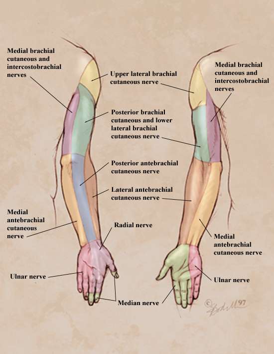
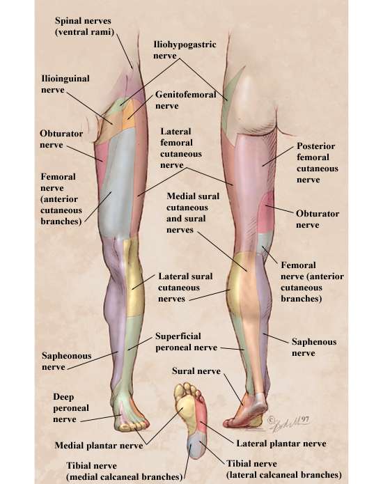
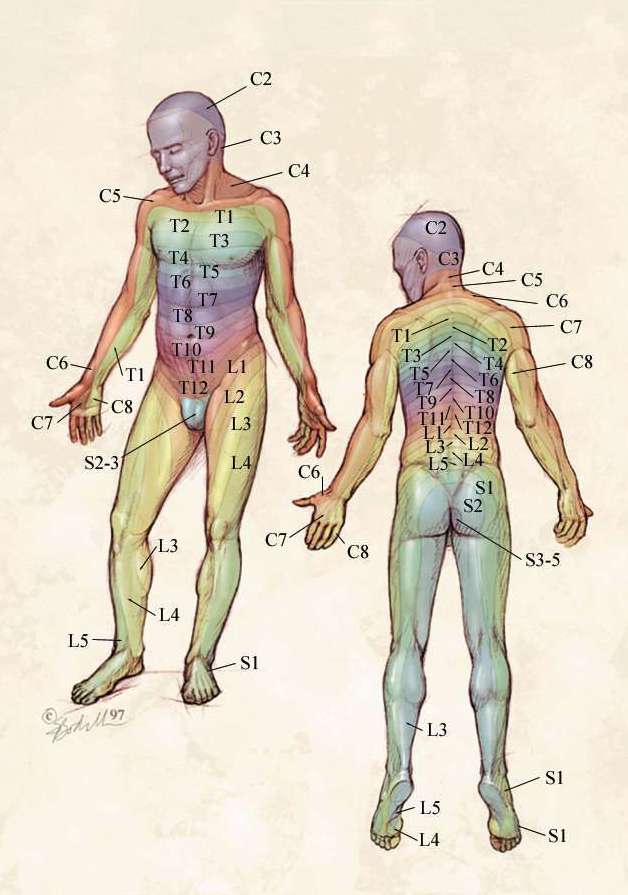
Dermatomes and peripheral nerves' cutaneous distribution.


Sensory dermatomes


Cutaneous nerve distribution of the upper limb


Cutaneous nerve distribution of the lower limb

Friday, 26 July 2013

Can you define health?


Can you define health? OK, we all know the definition of health by the World Health Organization. And I totally agree with this definition. But I want something more. I want something that I can apply to my self in practical terms as well to people that I am trying to treat.

The last 4-5 years in my life I came across to an excellent definition of health by a medical doctor specializing in Homeopathy. Dr. George Vithoulkas (introduced to me by Dr. Spyros Kyvelos) gives an excellent definition for health, in my opinion. You should read his article here in order to completely understand his point of view.

I always keep in my mind the following major points:

Health is:

- freedom from pain at the somatic level
- freedom from lust at the emotional level
- freedom from selfishness at the mental level

And a great way to measure health is the degree of creativity of the individual.

As Dr. Vithoulkas says "these are rules that nobody can escape from, whether he likes it or not". And I feel blessed that I met this man and his point of view on medicine as a science.

Thursday, 25 July 2013

The Resource - Physical Therapy Modalities Manual


A great modalities manual that explains in simple terms all the modalities used in Physical Therapy. It is true that these modalities are losing ground against manual therapy, kinesiotherapy and other more manual techniques, but it would be wrong to completely deny their use and effect on the human body. Anyhow, a good Physical Therapists should have all the possible tools in his arsenal and use each one when appropriate. Not every body is the same and or responds in the same way to every modality. Sp, the challenge for the Physical Therapist is to recognise and identify which tool can be effective is each case. 

Wednesday, 24 July 2013

Proper bike posture


With biking becoming more and more famous all over Europe, it is extremely important to know how to properly set your posture.

Interactive body


Interactive Body by moveformardpt.com.

Browse by body part to learn how a physical therapist can help you.

Why you should care about the drugs your Doctor prescribes


Watch the video and you will be amazed about this great project. They have taken the data and put it into an online database that allows anyone to look up a doctor's prescribing patterns and see how they compare with those of other doctors!!

Read also this very interesting article on this subject. it gives you a brief overview on the reasons you should care about the drugs your Doctor prescribes.

Neural tissue management


We all learn during our education the Upper and Lower Limps Tension Tests. These tests can be used also as mobilization and treatment techniques.

I recently read a few articles on the usefulness and the effect of these and some similar techniques on nerve related pain in the neck and the limps.

Here you can find a great Randomized Control Trial by the School of Health and Rehabilitation Sciences at the University of Queensland, Australia.

Here ypu have fine a novel protocol to develop a prediction model that identifies patients with nerve-related neck and arm pain who benefit from the early introduction of neural tissue management.

Here you can find a very interesting blog entry on neural tissue management.

Finally, here you can find a treatment approach for carpal tunnel syndrome suggested by http://www.handsonemg.com.

Tuesday, 23 July 2013

Can Physical Therapy help Angelina Jolie?


We all heard recently the story of Angelina Jolie and her decision to have a double mastectomy operation in order to minimize the possibilities to have breast cancer.

Did you know that Physical Therapy is recommended after this kind of operation? Dr. Dimitrios Kostopoulos co-founder of www.handsonpt.org is giving a brief talk on why this is true.

AlterG - the anti-gravity treadmill


The video presents a range of patients with different diagnoses using the AlterG - the anti-gravity treadmill:

1. A Parkinson's patient walking safely, allowing her to stay mobile and improve her condition
2. An overweight patient enjoying running for the first time and losing weight
3. A triathlete recovering from an injury and competing again
4. An injured cop recovering from an injury, enabling him to return to his job

I just came across this product. It seems to me that it would benefit these kinds of patients since it allows pain free movements that they would otherwise not be able to do - a fact good for both the musculoskeletal and the neurologic rehab process. This fits very well with the functional rehabilitation a Physical Therapist should focus on.

Monday, 22 July 2013

Biomechanics of skeletal muscle


A quick reference to the basic biomechanics of skeletal muscle.

Diagnostic imaging for Physical Therapists


The famous physio-pedia.com has created a wonderful article on diagnostic imaging for Physical Therapists. Read it here.

Meniscal tears


A physical therapists will face these situations quite often in his daily practise. It is useful to know the main types of a meniscal tear and base his treatment plan accordingly.

Wednesday, 17 July 2013

NSCA certification for fitness and training


For those Physical Therapists that are interested in focusing in Fitness and Training, I highly recommend this certification.

"NSCA is the worldwide authority on strength and conditioning, that supports and disseminates research-based knowledge and its practical application to improve athletic performance and fitness.

Our History

Founded in 1978, the NSCA has been serving its members by bridging the gap between science and application. Take an online tour of the NSCA’s legacy and learn about the association’s impact on strength and conditioning as both a practice and profession".

Monday, 15 July 2013

MovingNaturally Training


When I watched this video I liked very much this idea of regaining our abilities to move naturally. It is true that we leave a life that does not favor natural movement and restricts our body's amazing capabilities. Of course, there are a lot of ways you can follow to train them, but I liked this approach by MovNat.

The Functional Movement Screen


The Functional Movement Screen (FMS) is the product of an exercise philosophy known as Functional Movement Systems. This exercise philosophy and corresponding set of resources is based on sound science, years of innovation, and current research.

» How it Works - Simplifying Movement

Put simply, the FMS is a ranking and grading system that documents movement patterns that are key to normal function. By screening these patterns, the FMS readily identifies functional limitations and asymmetries. These are issues that can reduce the effects of functional training and physical conditioning and distort body awareness.

The FMS generates the Functional Movement Screen Score, which is used to target problems and track progress. This scoring system is directly linked to the most beneficial corrective exercises to restore mechanically sound movement patterns.

Exercise professionals monitor the FMS score to track progress and to identify those exercises that will be most effective to restore proper movement and build strength in each individual.

» What it Does - Widespread Benefits

The FMS simplifies the concept of movement and its impact on the body. Its streamlined system has benefits for everyone involved - individuals, exercise professionals, and physicians.

Communication - The FMS utilizes simple language, making it easy for individuals, exercise professionals, and physicians to communicate clearly about progress and treatment.

Evaluation - The screen effortlessly identifies asymmetries and limitations, diminishing the need for extensive testing and analysis.

Standardization - The FMS creates a functional baseline to mark progress and provides a means to measure performance.

Safety - The FMS quickly identifies dangerous movement patterns so that they can be addressed. It also indicates an individual’s readiness to perform exercise so that realistic goals can be set and achieved.

Corrective Strategies - The FMS can be applied at any fitness level, simplifying corrective strategies of a wide array of movement issues. It identifies specific exercises based on individual FMS scores to instantly create customized treatment plans.

The Mulligan concept


Manual Therapy – NAGS, SNAGS, MWMs | Mulligan Concept

Mobilization with Movement manual therapy techniques were discovered and developed by Brian Mulligan F.N.Z.S.P. (Hon), Dip. M.T., Wellington, New Zealand. This simple yet effective manual approach addresses musculoskeletal disorders with pain free manual joint “repositioning” techniques for restoration of function and abolition of pain.

Explore this Official International Web site to find more about the Mulligan Concept.

Master Programme in Sport Science, Sports Medicine


Lund University in Sweden:


Master's Programme, 2 Years, Full time
Code: LU-18520
120 credits
Lund Campus
Study period: 02 Sep 2013 – 07 Jun 2015
The aim of the programme is to provide in-depth knowledge, skills and abilities in the field of sport sciences, from a number of different scientific perspectives. Particular focus is placed on advanced in-depth study of sports medicine and sport psychology.

The programme includes second-cycle courses in

  • prevention and treatment of sports injuries
  • sports nutrition
  • exercise physiology
  • leadership and communication
  • mental aspects of sport and physical performance
  • applied sport and exercise psychology (team and individual)
  • An important aspect of the programme is close contact with potential future employers and workplaces through internships.

Saturday, 13 July 2013

Clinical guidelines for stroke management


The National Stroke Foundation: We are a not-for-profit organisation that works with stroke survivors, carers, health professionals, government and the public to reduce the impact of stroke on the Australian community.

We are the voice of stroke in Australia. Our mission is to stop stroke, save lives and end suffering.

Here is the clinical guidelines they have developed for stroke management.

I have already posted the KNGF's guidelines for stroke patients, too.

Clinical practice guidelines for the management of rotator cuff syndrome in the workplace


The University of New South Wales Rural Clinical School, Port Macquarie has developed guidelines for the clinical management of rotator cuff syndrome in the workplace. Shoulder pain is a common musculoskeletal presentation in primary care practice – both degenerative and acute. As such, it provides a challenge to all involved in prevention and treatment, from patients to clinicians to employers. The primary objective of these guidelines is to provide recommendations, based on current evidence, which will hopefully improve clinical outcomes for workers, employers and health care providers.

Friday, 12 July 2013

Medbridge education online


MedBridge Education


Learn, Connect, Succeed

MedBridge Education dedicates itself to providing high quality online continuing education for Physical Therapy professionals. By collaborating with top-tier clinicians and technology-learning integration experts, MedBridge Education has created a premier curriculum designed specifically for online delivery. You will learn up-to-date, evidence-based curriculum, and proven rehabilitation techniques designed to immediately help your patients.

Deep brain stimulation for Parkinson's disease


Deep brain stimulation (DBS) is a surgical procedure used to treat a variety of disabling neurological symptoms—most commonly the debilitating symptoms of Parkinson’s disease (PD), such as tremor, rigidity, stiffness, slowed movement, and walking problems.  The procedure is also used to treat essential tremor, a common neurological movement disorder.  At present, the procedure is used only for patients whose symptoms cannot be adequately controlled with medications.

DBS uses a surgically implanted, battery-operated medical device called a neurostimulator—similar to a heart pacemaker and approximately the size of a stopwatch—to deliver electrical stimulation to targeted areas in the brain that control movement, blocking the abnormal nerve signals that cause tremor and PD symptoms.

Barium swallow and achalasia


A barium swallow (or esophagography) is a medical imaging procedure used to examine the upper GI (gastrointestinal) tract, which includes the esophagus and, to a lesser extent, the stomach.

Achalasia, also known as esophageal achalasia, achalasia cardiae, cardiospasm, and esophageal aperistalsis, is an esophageal motility disorder involving the smooth muscle layer of the esophagus and the lower esophageal sphincter (LES). It is characterized by incomplete LES relaxation, increased LES tone, and lack of peristalsis of the esophagus (inability of smooth muscle to move food down the esophagus) in the absence of other explanations like cancer or fibrosis.

Inspired by orthopedicmanualpt.com.

Differential diagnosis: femoroacetabular impingement


Orthopedic Manual Physical Therapy

A very useful article on femoroacetabular impingement by orthopedicmanualpt.com. Read it all and keep in mind that

...treatment and diagnosis of femoroacetabular impingement is in its infancy, however by utilizing current evidence and relying on all aspects of your patient’s evaluation and response to treatment, your patient outcomes will continue to improve as our knowledge of this condition continues to grow.

...interventions found to be beneficial included joint mobilization (Long-axis Distraction, Lateral distraction, anterior glide, and posterior glide), core strengthening, gluteal strengthening, proprioception, and hip flexor stretching. Additionally, several studies found that passive range of motion of the hip results in counter-productive exacerbation of symptoms and should be avoided in lieu of the aforementioned beneficial interventions.

Mechanisms of somatic pain


This article, by the University of Utah pain research center, is very interesting but a little bit too detailed. But is worths reading it once and keeping in mind the  summary points:

  1. Somatic pain is normally triggered by the activation of nociceptors. Particular types of nociceptors have been well characterized in cutaneous, articular and muscle nerves.
  2. The activation of cutaneous Ad nociceptors causes a sensation of pricking pain, whereas stimulation of C polymodal nociceptors elicits burning pain. Muscle nociceptors produce aching pain.
  3. Unlike sensitive mechanoreceptors and thermoreceptors, nociceptors can be sensitized by damaging stimuli. Sensitization appears to be triggered by the release of chemical substances, such as prostaglandins, bradykinin, serotonin, and histamine, into the environment of peripheral nociceptor terminals. Some nociceptors are quite unresponsive until they are sensitized.
  4. Nociceptors project to particular laminae in the spinal cord dorsal horn. Cutaneous Ad nociceptive fibers end in laminae I, II, and V, whereas cutaneous C polymodal nociceptors end chiefly in lamina II. Fiber, joint, and muscle afferents project to laminae I and V.
  5. Fine afferent terminals, presumably of nociceptors, in the dorsal horn contain peptides, such as substance P and CGRP, and also excitatory amino acids. Both classes of substances are likely to be released during intense noxious stimulation.
  6. Noxious stimuli trigger both excitatory and inhibitory events in the dorsal horn. Inhibition is likely to be mediated by such agents as inhibitory amino acids and inhibitory peptides. The circuits may be local or involve a supraspinal loop.
  7. STT cells that project to the ventral posterior lateral thalamic nucleus in monkeys and rats have response properties that suit them for a role in the sensory-discriminative aspects of pain. Their input can be from cutaneous, articular, muscle and/or visceral receptors. Convergent inputs may account for pain referral.
  8. The responses of STT cells are altered by pathological processes. These neurons become more responsive following damage of the skin by intense mechanical, thermal, or chemical stimuli. A similar change occurs during the development of experimental acute arthritis. It is proposed that sensitization of STT cells helps account for the development of primary and secondary hyperalgesia and allodynia following damage.
  9. The mechanism of sensitization of STT cells is likely to involve excitatory amino acid and NK1 receptors.
  10. Experimental models of painful neuropathy are being developed by several groups. The responses of STT cells in these models are altered in a fashion consistent with the development of spontaneous pain, allodynia, and hyperalgesia.

The spinal cord and spinal tracts by Professor Fink


In Part 1 of Professor Fink's 2-Part Series on the Spinal Cord, he reviews the anatomy of the Spinal Cord and the functional organization at each segmental level. Professor Fink describes the horizontal flow of sensory information into the Spinal Cord and the flow of motor commands out of the Spinal Cord. Reference is made to Gray Matter, White Matter, Spinal Nerves, Dorsal Root Ganglion, Ventral Root, Commissures, decussation, Somatic Reflexes, Dorsal (Posterior) Gray Horn, Ventral (Anterior) Gray Horn, Lateral Gray Horn.


In Part 2 of Professor Fink's 2-Part Series on the Spinal Cord, he reviews the anatomy of the Sensory and Motor Tracts located in the White Matter of the Spinal Cord, describing the transmission of signals vertically within the CNS. Reference is made to the Spinothalamic Tract, Dorsal White Columns (Fasciculus Gracilis & Fasciculus Cuneatus), Spinocerebellar Tract, Corticospinal (Pyramidal) Tract, Extracorticospinal (Extrapyramidal) Tract, Spinal Cord Injuries (Lesions), Herniated Disk, paralysis.

You can also read my own work on the most basic material you need to know on neurology as a Physical therapy student.

Robert Gordon University, Aberdeen


"With inter-professional working ever more important, our strong focus in this area is a big ‘plus' post-graduation. We promote hands-on, practical, patient-centred training that leads to professional registration through our undergraduate courses and high quality care.

We're responsive to health and social care policy, also providing a service to the local community while training - for example through local volunteer patients.

You'll be taught by experienced practitioner staff who are also active researchers in their field. Strong links with practice colleagues ensure up-to-date courses that equip our graduates to enter the workplace with specific skills attractive to domestic and international markets. You'll also learn transferable skills in communication and team working.

Professional placements throughout many courses start as early as year one - earlier than at some other universities. This allows students to start putting theory into practice as soon as possible".

The McKenzie Method


Robin Anthony McKenzie

The McKenzie Method is not merely extension exercises. In its truest sense, McKenzie is a comprehensive approach to the spine based on sound principles and fundamentals that when understood and followed accordingly are very successful. In fact, most remarkable, but least appreciated, is the McKenzie assessment process.

Assessment. Unique to the McKenzie Method is a well-defined algorithm that leads to the simple classification of spinal-related disorders. It is based on a consistent "cause and effect" relationship between historical pain behavior as well as the pain response to repeated test movements, positions and activities during the assessment process.

A systematic progression of applied mechanical forces (the cause) utilizes pain response (the effect) to monitor changes in motion/function. The underlying disorder can then be quickly identified through objective findings for each individual patient. The McKenzie classification of spinal pain provides reproducible means of separating patients with apparently similar presentations into definable sub-groups (syndromes) to determine appropriate treatment.

McKenzie has named these three mechanical syndromes: Postural, Dysfunction and Derangement.

Postural: End-range stress of normal structures
Dysfunction: End-range stress of shortened structures (scarring, fibrosis, n.root adherence)
Derangement: Anatomical disruption or displacement within the motion segment
(All three mechanical syndromes – postural, dysfunction, and derangement – occur in the cervical as well as thoracic and lumbar regions of the spine.)

Thursday, 11 July 2013

Kevin Stone: The bio-future of joint replacement


Arthritis and injury grind down millions of joints, but few get the best remedy -- real biological tissue. Kevin Stone shows a treatment that could sidestep the high costs and donor shortfall of human-to-human transplants with a novel use of animal tissue.

Inspired by www.thestudentphysicaltherapist.com.

Monday, 8 July 2013

Physical Therapist: 8th best career, 5th best health care job


According to money.usnews.com, the career of a Physical Therapist is ranked 8th in the 100 best careers. Besides that, it is ranked 5th in the best healthcare jobs:

"Today our focus on preventing disease, illness, and injury is just as apparent as our fight to diagnose and treat them. And as even our youngest baby boomers grow closer to age 50, the need to employ qualified healthcare personnel to both prevent and treat medical conditions intensifies. The Bureau of Labor Statistics (BLS) reports that we not only need to retain those workers already in the field, but also add a substantial number of new ones, with the most occupational growth expected among healthcare support jobs. That's why for this year's list of Best Healthcare Jobs, we included the usual suspects, like nurses and physicians, but also a few unexpected picks, like diagnostic medical sonographer and veterinary tech".

Interesting also to read about the U.S. News best jobs rankings methodology.

Friday, 5 July 2013

Having a short Achilles tendon - can you benefit from it?


An interesting article on achilles tendon length.

Having a short Achilles tendon may be an athlete's Achilles heel

Tendon length varies in all major groups and from person to person. Tendon length is practically the discerning factor where muscle size and potential muscle size is concerned. For example, should all other relevant biological factors be equal, a man with a shorter tendons and a longer biceps muscle will have greater potential for muscle mass than a man with a longer tendon and a shorter muscle. Successful bodybuilders will generally have shorter tendons. Conversely, in sports requiring athletes to excel in actions such as running or jumping, it is beneficial to have longer than average Achilles tendon and a shorter calf muscle.

Pelvic floor muscles physiology


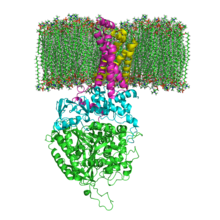
Succinate Dehydrogenase 1YQ3 and Membrane.png

I recently read an research article on pelvic floor muscle assessment and I found interesting the following parts:
  1. The pelvic floor muscles (PFM) consist of approximately 70% slow-twitch (type 1) and 30% fast-twitch (type 2) muscle fibres (Gilpin et al, 1989).
  2. During a vaginal examination using the distal pad of the index finger to palpate the perivaginal muscles, a definite bulging and lifting of the muscles are felt during a contraction, and in both the contracted and relaxed state, areas of atrophy can be detected.
  3. A moderate to strong contraction of the levator ani muscles has both a squeeze and a lift component.
  4. Regarding different muscle components (that is, fast- and slow-twitch fibres), it has been shown that fast fibres are recruited only during activities involving speed and/or power (Edwards, 1978) and if the response to a command to contract the PFM maximally is sluggish, then it is probable that only slow-twitch muscle fibres are being used. Jones and Rutherford (1987) reported that in some untrained muscles, the fast-firing muscle fibres are never recruited, and so these may need targeting in some other way during PFM rehabilitation. Gosling et al (1981) described the PFM slow-twitch fibres as being responsible for maintaining continuous muscle activity over prolonged periods, with the fast-twitch fibres recruited reflexly during sudden increases in intra-abdominal pressure, for example when coughing. A reduction in slow-twitch activity would therefore manifest itself in a short duration contraction and few repetitions, and PFM in this category may thus be responsible for decreased support of the proximal urethra and reduced urethral occlusion. A reduction in fast-twitch fibre activity may result in a slower, weaker reflex response to increases in intra-abdominal pressure. Logically, one should assess both the fast- and slow-twitch muscle fibre activity before planning an exercise regimen.
  5. Exercise programme planning: the plethora of PFM exercise regimens in the literature indicates a lack of standardisation and, when compared with general rehabilitation methodology, the reports show lack of scientific application. There is a wide range of muscular strength and endurance across any female population, and so it is postulated that a uniform, standard regimen is not appropriate. Instead, assessment of the fast- and slow-twitch components, leading to an individual exercise programme, has been proposed, to target specifically the weakness of each individual patient.
  6. Muscle weakness leads to reduction in mitochondrial oxidative capacity as indicated by a decrease in the succinate dehydrogenase (SDH) activity (Eriksson and Haggmark, 1979). Consequently, these same authors maintain that it is necessary to gradually increase the SDH reserves by repeated contractions which will deplete levels and stimulate greater production. This reinforces the theory of overload and supports the need for a programme of regular daily contractions advanced in this study.

Interesting also to know that SDH or succinate-coenzyme Q reductase (SQR) or respiratory Complex II is an enzyme complex, bound to the inner mitochondrial membrane of mammalian mitochondria and many bacterial cells. It is the only enzyme that participates in both the citric acid cycle and the electron transport chain.

The fundamental role of SDH in the electron transfer chain of mitochondria makes it vital in most multicellular organisms, removal of this enzyme from the genome has also been shown to be lethal at the embryonic stage in mice. Mutations of different genes used in its construction can lead to:
  • Leigh syndrome, mitochondrial encephalopathy, and optic atrophy
  • Tumorogenesis in chromaffin cells, causing hereditary paraganglioma and hereditary pheochromocytoma. Tumors tend to be malignant. It can also lead to decreased life-span and increased production of superoxide ions.
  • Decreased life-span, increased production of superoxide ions, hereditary paraganglioma and hereditary pheochromocytoma. Tumors tend to be benign. These mutations are uncommon.
  • Hereditary paraganglioma and hereditary pheochromocytoma. Tumors tend to be benign, and occur often in the head and neck regions. These mutations can also decrease life-span and increase production of superoxide ions.

Mammilian succinate dehydrogenase functions not only in mitochondrial energy generation, but also has a role in oxygen sensing and tumor suppression; and, therefore, is the object of ongoing research.

Wednesday, 3 July 2013

The American Academy of Orthopaedic Surgeons


AAOS: American Academy of Orthopaedic Surgeons® / American Association of Orthopaedic Surgeons®


The American Academy of Orthopaedic Surgeons

Founded in 1933, the Academy is the preeminent provider of musculoskeletal education to orthopaedic surgeons and others in the world. Its continuing medical education activities include a world-renowned Annual Meeting, multiple CME courses held around the country and at the Orthopaedic Learning Center, and various medical and scientific publications and electronic media materials.

The American Association of Orthopaedic Surgeons

Founded by the Academy Board of Directors in 1997, the Association engages in health policy and advocacy activities on behalf of musculoskeletal patients and the profession of orthopaedic surgery.

A traditional symbol of orthopaedics is the bent tree that has been braced to make it grow straight.

Why we call it "orthopaedics"
Since orthopaedics' beginnings, its specialists have treated children suffering from spine and limb deformities. The Greek roots of the word "orthopaedics" are ortho (straight) and pais (child). Early orthopaedists often used braces or other forms of treatment to make the child "straigh.

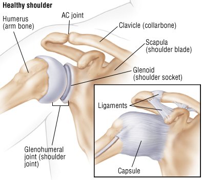
Shoulder disclocation


Anterior (forward)

Anterior dislocations are usually caused by a direct blow to or fall on an outstretched arm. The patient typically appears holding their arm externally rotated and slightly abducted.
Posterior (backward)

Posterior dislocations are occasionally due to electric shock or seizure and may be caused by strength imbalance of the rotator cuff muscles. Patients typically present holding their arm internally rotated and adducted, and exhibiting flattening of the anterior shoulder and a prominent coracoid process.
Inferior (downward)

Inferior dislocation is the least likely form, occurring in less than 1% of all shoulder dislocation cases. This condition is also called luxatio erecta because the arm appears to be permanently held upward or behind the head. It is caused by a hyper abduction of the arm that forces the humeral head against the acromion.

Golgi tendon reflex vs. stretch reflex


Two different reflexes exist in skeletal muscles: the Golgi tendon reflex and the stretch reflex. It is very important for a Physical Therapist to understand the structure and function of these two reflexes, because he will use their effect on muscles in many different cases.

In the conceptual model above, I have drawn an agonist muscle (the big orange cylinder), an antagonist muscle (the smaller orange cylinder) and a spinal segment (the grey region). In the agonist muscle I have also drawn two sensory organs: the Golgi tendon organ and the muscle spindle.

The Golgi tendon organ senses changes in muscle tension. It is a proprioceptive sensory receptor organ that is located at the origins and insertion of skeletal muscle fibers into the tendons of skeletal muscle. It provides the sensory component of the Golgi tendon reflex. In a Golgi tendon reflex, skeletal muscle contraction causes the agonist muscle to simultaneously lengthen and relax. Though muscle tension is increasing during the contraction, alpha motor neurons in the spinal cord supplying the muscle are inhibited through the Inhibitory Postsynaptic Potential. However, antagonistic muscles are activated.

An inhibitory postsynaptic potential (IPSP) is a kind of synaptic potential that makes a postsynaptic neuron less likely to generate an action potential. The opposite of an inhibitory postsynaptic potential is an excitatory postsynaptic potential (EPSP), which is a synaptic potential that makes a postsynaptic neuron more likely to generate an action potential. They can take place at all chemical synapses which use the secretion of neurotransmitters to create cell to cell signalling. Inhibitory presynaptic neurons release neurotransmitters which then bind to the postsynaptic receptors; this induces a postsynaptic conductance change as ion channels open or close. An electrical current is generated which changes the postsynaptic membrane potential to create a more negative postsynaptic potential.

Muscle spindles are sensory receptors within the belly of a muscle, which primarily detect changes in the length of this muscle. They convey length information to the central nervous system via sensory neurons. This information can be processed by the brain to determine the position of body parts. The responses of muscle spindles to changes in length also play an important role in regulating the contraction of muscles, by activating motoneurons via the stretch reflex to resist muscle stretch. The stretch reflex (myotatic reflex) is a muscle contraction in response to stretching within the muscle. It is a monosynaptic reflex which provides automatic regulation of skeletal muscle length.

When a muscle lengthens, the muscle spindle is stretched and its nerve activity increases. This increases alpha motor neuron activity, causing the muscle fibers to contract and thus resist the stretching. A secondary set of neurons also causes the opposing muscle to relax. The reflex functions to maintain the muscle at a constant length.

Tuesday, 2 July 2013

Proximal tibiofibular ligament instability


A nice and simple article by Dr. Robert F. LaPrade on this very rare pathology.

Injury to the proximal tibiofibular joint is rather rare, but it can be debilitating in patients who have symptoms.  The usual mechanism is having a patient fall on a plantar-flexed ankle, with the stress being brought through the fibula, such that the proximal fibula will sublux out of place over the lateral aspect of the knee joint.  In other circumstances, there may be a more obvious injury where one has a significant trauma or motor vehicle accident where the proximal tibiofibular joint is disrupted. 

In most circumstances, it is the posterior proximal tibiofibular joint ligament which is disrupted.  This results in the fibula rotating away from the tibia during deep squatting.  This can result in a feeling of instability, a visible bony deformity, and concurrent irritation of the common peroneal nerve.  This is because the common peroneal neve crosses the lateral aspect of the fibular neck within 2-3 cm of the lateral aspect of the fibular head. 


Selected as "one of the Best Doctors in America", Dr. Robert F. LaPrade is a complex orthopaedic (orthopedic) knee surgeon at The Steadman Clinic in Vail, CO specializing in complex knee injuries.  He also serves as Chief Medical Officer, Deputy Director of the Sports Medicine Fellowship Program and Director of the International Research Scholar Program at the Steadman Philippon Research Institute.

Monday, 1 July 2013

The anatomical snuff box


This small cavity that appears at the radial side of the hand when we fully extend and abduct our thumb is call "anatomical snuff box" or "tabatiere anatomique".

The bony borders of this cavity are:

(bottom surface) the trapezium and scaphoideum
(proximally) the styloid process of the radius
(distally) the apex of the muscle tendons triangle

Three tendons define the borders of this cavity, as seen in the picture above:

1. Extensor pollicis longus

Origin: dorsal surface of the ulnar & the membrana interossea
Insertion: phalanx I (dorsally at the base of the distal phalanx)
Action: wrist: RAD DIV, EXT - thump: EXT at the metacarpophalangeal and the interphalangeal joints
While abductor pollicis brevis and adductor pollicis, both attached to the extensor pollicis longus tendon, can extend the thumb's interphalangeal joint to the neutral position, only extensor pollicis longus can achieve full hyperextension at the interphalangeal joint
Innervation: n. Radialis (C7-C8)


2. Extensor pollicis brevis

Origin: dorsal surface of the radius & the membrana interossea
Insertion: phalanx I (dorsally at the base of the proximal phalanx)
Action: wrist: RAD DIV - thump: EXT at the carpometacarpal and the metacarpophalangeal joints
Innervation: n. Radialis (C7-C8)


3. Abductor pollicis longus

Origin: dorsal surface of the ulnar & the radius & the membrana interossea
Insertion: base of the metacarpi I
Action: wrist: RAD DIV - thump: ABD at the carpometacarpal joint
Innervation: n. Radialis (C7-C8)


Besides, the radial artery of the forearm passes through this cavity. It runs distally on the anterior part of the forearm. There, it serves as a landmark for the division between the anterior and posterior compartments of the forearm, with the posterior compartment beginning just lateral to the artery. The artery winds laterally around the wrist, passing through the anatomical snuff box and between the heads of the first dorsal interosseous muscle.


The dorsal (superficial) cutaneous branch of the radial nerve can be palpated by stroking along the extensor pollicis longus with the dorsal aspect of a fingernail.

Pathology

1. Scaphoideum fracture

In the event of a fall onto an outstretched hand, the articulation between the scaphoideum and the radius is the area through which the brunt of the force will focus. In case the force is big enough the scaphoideum may fracture. There are two anatomical peculiarities that make this fracture quite dangerous and difficult to treat:

a. The size of the scaphoideum - the scaphoid is a small, oddly shaped bone whose purpose is to facilitate mobility rather than confer stability to the wrist joint. In the event of inordinate application of force over the wrist, this small scaphoid is clearly likely to be the weak link.

b. The vascularization of the scaphoideum - blood enters the scaphoid distally. Consequently, in the event of a fracture the proximal segment of the scaphoid will be devoid of a vascular supply, and will - if action is not taken - avascularly necrose within a sufferer's snuffbox.

2. DeQuervain’s tenosynovitis

DeQuervain's tenosynovitis is an inflammation of the fluid-filled sheath (called the synovium) that surrounds the tendons of the muscles mentioned above. Mostly, the extensor pollicis brevis and the abductor pollicis longus are the muscles affected in that case. The main reason for this inflammation is overuse syndromes. The main symptoms are pain, tenderness, and swelling over the thumb side of the wrist, and difficulty gripping. The Finkelstein's test may be positive.

3. Cheiralgia paresthetica

Cheiralgia paresthetica or Wartenberg's syndrome is the compression of the radial nerve along its course in the forearm. The area affected is typically on the back or side of the hand at the base of the thumb, near the anatomical snuffbox, but may extend up the back of the thumb and index finger and across the back of the hand. The most common cause is thought to be constriction of the wrist, as with a bracelet or watchband. Symptoms include numbness, tingling, burning or pain. Since the nerve branch is sensory there is no motor impairment.

There might be other injuries that can cause symptoms around the region as well like osteoarthritis, carpal instabilities, or cervical radiculopathy (C7-8). However, these were the 3 main problems that can appear and a Physical Therapist should at least be aware of in order to successfully assess a patient.

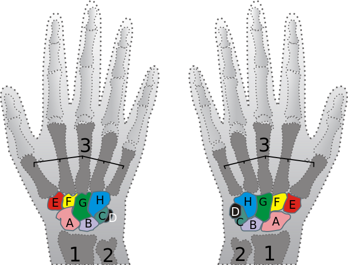
The bones of the carpus


File:Carpus.svg

Difficult to learn them, but it can become easy if you use these pictures:

Picture 1 (left) - view from below
Picture 2 (right) - view from below

A: Scaphoideum, B: Lunate, C: Triquetrum, D: Prisiform, E: Trapezium, F: Trapezoideum,
G: Capitatum, H: Hamatum

A file I have created with all the muscles of the upper limbs is here.

The bones of the tarsus


File:Tarsus.png

Difficult to learn them, but it can become easy if you use these pictures:

Picture 1 (left) - view from below
Picture 2 (right) - view from below

A: Calcaneus, B: Talus, C: Cuboid, D: Navicular, E-F-G: 1st, 2nd, 3rd Cuneiform

A file I have created with all the muscles of the lower limbs is here.